Magnesium »
PDB 4ko8-4kvw »
4kva »
Magnesium in PDB 4kva: Gtpase Domain of Septin 10 From Schistosoma Mansoni in Complex with Gtp
Protein crystallography data
The structure of Gtpase Domain of Septin 10 From Schistosoma Mansoni in Complex with Gtp, PDB code: 4kva
was solved by
A.E.Zeraik,
H.M.Pereira,
Y.V.Santos,
J.Brandao-Neto,
R.C.Garratt,
A.P.U.Araujo,
R.Demarco,
with X-Ray Crystallography technique. A brief refinement statistics is given in the table below:
| Resolution Low / High (Å) | 74.22 / 2.14 |
| Space group | C 1 2 1 |
| Cell size a, b, c (Å), α, β, γ (°) | 160.800, 47.040, 95.480, 90.00, 112.61, 90.00 |
| R / Rfree (%) | 18.7 / 22.1 |
Magnesium Binding Sites:
The binding sites of Magnesium atom in the Gtpase Domain of Septin 10 From Schistosoma Mansoni in Complex with Gtp
(pdb code 4kva). This binding sites where shown within
5.0 Angstroms radius around Magnesium atom.
In total 2 binding sites of Magnesium where determined in the Gtpase Domain of Septin 10 From Schistosoma Mansoni in Complex with Gtp, PDB code: 4kva:
Jump to Magnesium binding site number: 1; 2;
In total 2 binding sites of Magnesium where determined in the Gtpase Domain of Septin 10 From Schistosoma Mansoni in Complex with Gtp, PDB code: 4kva:
Jump to Magnesium binding site number: 1; 2;
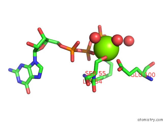
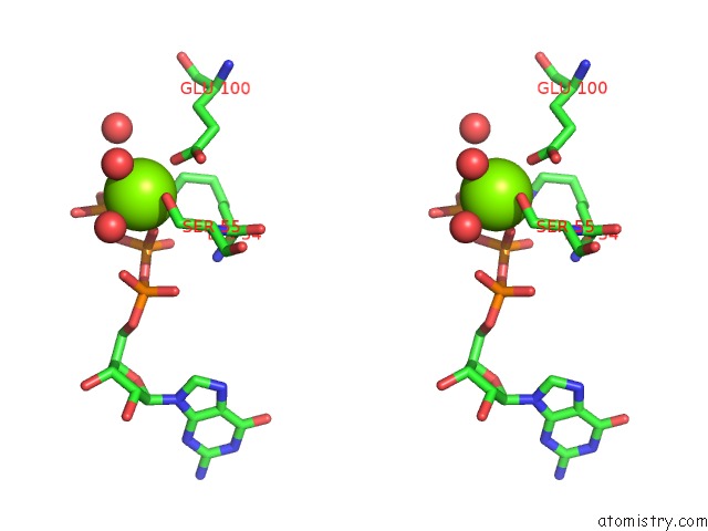
Magnesium binding site 1 out of 2 in 4kva
Go back to
Magnesium binding site 1 out
of 2 in the Gtpase Domain of Septin 10 From Schistosoma Mansoni in Complex with Gtp

Mono view

Stereo pair view
Mono view
Stereo pair view
A full contact list of Magnesium with other atoms in the Mg binding
site number 1 of Gtpase Domain of Septin 10 From Schistosoma Mansoni in Complex with Gtp within 5.0Å range:
|
Magnesium binding site 2 out of 2 in 4kva
Go back to
Magnesium binding site 2 out
of 2 in the Gtpase Domain of Septin 10 From Schistosoma Mansoni in Complex with Gtp
Mono view
Stereo pair view
Mono view
Stereo pair view
A full contact list of Magnesium with other atoms in the Mg binding
site number 2 of Gtpase Domain of Septin 10 From Schistosoma Mansoni in Complex with Gtp within 5.0Å range:
|
Reference:
A.E.Zeraik,
H.M.Pereira,
Y.V.Santos,
J.Brandao-Neto,
M.Spoerner,
M.S.Santos,
L.A.Colnago,
R.C.Garratt,
A.P.Araujo,
R.Demarco.
Crystal Structure of A Schistosoma Mansoni Septin Reveals the Phenomenon of Strand Slippage in Septins Dependent on the Nature of the Bound Nucleotide. J.Biol.Chem. V. 289 7799 2014.
ISSN: ISSN 0021-9258
PubMed: 24464615
DOI: 10.1074/JBC.M113.525352
Page generated: Sat Aug 17 03:59:19 2024
ISSN: ISSN 0021-9258
PubMed: 24464615
DOI: 10.1074/JBC.M113.525352
Last articles
Fe in 2YXOFe in 2YRS
Fe in 2YXC
Fe in 2YNM
Fe in 2YVJ
Fe in 2YP1
Fe in 2YU2
Fe in 2YU1
Fe in 2YQB
Fe in 2YOO