Magnesium »
PDB 4kvw-4l9z »
4l1k »
Magnesium in PDB 4l1k: Crystal Structure of D-Alanine-D-Alnine Ligase From Xanthomonas Oryzae Pv. Oryzae with Amppnp
Enzymatic activity of Crystal Structure of D-Alanine-D-Alnine Ligase From Xanthomonas Oryzae Pv. Oryzae with Amppnp
All present enzymatic activity of Crystal Structure of D-Alanine-D-Alnine Ligase From Xanthomonas Oryzae Pv. Oryzae with Amppnp:
6.3.2.4;
6.3.2.4;
Protein crystallography data
The structure of Crystal Structure of D-Alanine-D-Alnine Ligase From Xanthomonas Oryzae Pv. Oryzae with Amppnp, PDB code: 4l1k
was solved by
T.T.N.Doan,
J.K.Kim,
L.W.Kang,
with X-Ray Crystallography technique. A brief refinement statistics is given in the table below:
| Resolution Low / High (Å) | 31.59 / 2.30 |
| Space group | P 43 21 2 |
| Cell size a, b, c (Å), α, β, γ (°) | 83.018, 83.018, 97.364, 90.00, 90.00, 90.00 |
| R / Rfree (%) | 21.1 / 26.4 |
Magnesium Binding Sites:
The binding sites of Magnesium atom in the Crystal Structure of D-Alanine-D-Alnine Ligase From Xanthomonas Oryzae Pv. Oryzae with Amppnp
(pdb code 4l1k). This binding sites where shown within
5.0 Angstroms radius around Magnesium atom.
In total only one binding site of Magnesium was determined in the Crystal Structure of D-Alanine-D-Alnine Ligase From Xanthomonas Oryzae Pv. Oryzae with Amppnp, PDB code: 4l1k:
In total only one binding site of Magnesium was determined in the Crystal Structure of D-Alanine-D-Alnine Ligase From Xanthomonas Oryzae Pv. Oryzae with Amppnp, PDB code: 4l1k:
Magnesium binding site 1 out of 1 in 4l1k
Go back to
Magnesium binding site 1 out
of 1 in the Crystal Structure of D-Alanine-D-Alnine Ligase From Xanthomonas Oryzae Pv. Oryzae with Amppnp
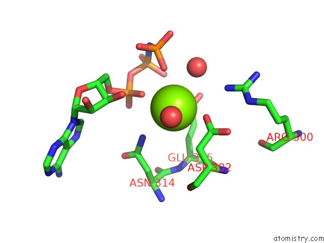
Mono view
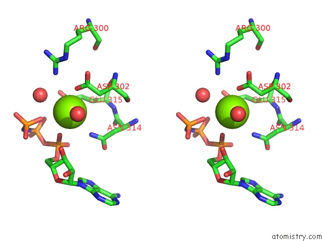
Stereo pair view
Mono view
Stereo pair view
A full contact list of Magnesium with other atoms in the Mg binding
site number 1 of Crystal Structure of D-Alanine-D-Alnine Ligase From Xanthomonas Oryzae Pv. Oryzae with Amppnp within 5.0Å range:
|
Reference:
T.T.N.Doan,
J.K.Kim,
H.P.T.Ngo,
H.T.Tran,
S.S.Cha,
K.M.Chung,
K.H.Huynh,
Y.J.Ahn,
L.W.Kang.
Crystal Structures of D-Alanine-D-Alanine Ligase From Xanthomonas Oryzae Pv. Oryzae Alone and in Complex with Nucleotides Arch.Biochem.Biophys. V.545C 92 2014.
ISSN: ISSN 0003-9861
PubMed: 24440607
DOI: 10.1016/J.ABB.2014.01.009
Page generated: Sat Aug 17 04:25:46 2024
ISSN: ISSN 0003-9861
PubMed: 24440607
DOI: 10.1016/J.ABB.2014.01.009
Last articles
Zn in 9J0NZn in 9J0O
Zn in 9J0P
Zn in 9FJX
Zn in 9EKB
Zn in 9C0F
Zn in 9CAH
Zn in 9CH0
Zn in 9CH3
Zn in 9CH1