Magnesium »
PDB 4kvw-4l9z »
4l6c »
Magnesium in PDB 4l6c: Crystal Structure of Human Mitochondrial Deoxyribonucleotidase in Complex with the Inhibitor Pib-T
Protein crystallography data
The structure of Crystal Structure of Human Mitochondrial Deoxyribonucleotidase in Complex with the Inhibitor Pib-T, PDB code: 4l6c
was solved by
P.Pachl,
J.Brynda,
P.Rezacova,
with X-Ray Crystallography technique. A brief refinement statistics is given in the table below:
| Resolution Low / High (Å) | 34.84 / 1.80 |
| Space group | P 43 21 2 |
| Cell size a, b, c (Å), α, β, γ (°) | 73.810, 73.810, 105.510, 90.00, 90.00, 90.00 |
| R / Rfree (%) | 15 / 17.2 |
Other elements in 4l6c:
The structure of Crystal Structure of Human Mitochondrial Deoxyribonucleotidase in Complex with the Inhibitor Pib-T also contains other interesting chemical elements:
| Iodine | (I) | 1 atom |
Magnesium Binding Sites:
The binding sites of Magnesium atom in the Crystal Structure of Human Mitochondrial Deoxyribonucleotidase in Complex with the Inhibitor Pib-T
(pdb code 4l6c). This binding sites where shown within
5.0 Angstroms radius around Magnesium atom.
In total only one binding site of Magnesium was determined in the Crystal Structure of Human Mitochondrial Deoxyribonucleotidase in Complex with the Inhibitor Pib-T, PDB code: 4l6c:
In total only one binding site of Magnesium was determined in the Crystal Structure of Human Mitochondrial Deoxyribonucleotidase in Complex with the Inhibitor Pib-T, PDB code: 4l6c:
Magnesium binding site 1 out of 1 in 4l6c
Go back to
Magnesium binding site 1 out
of 1 in the Crystal Structure of Human Mitochondrial Deoxyribonucleotidase in Complex with the Inhibitor Pib-T
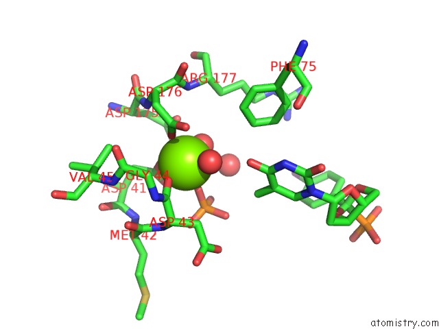
Mono view
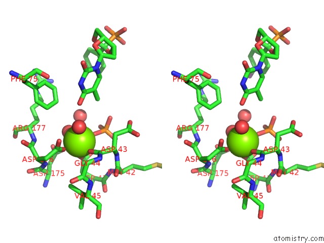
Stereo pair view
Mono view
Stereo pair view
A full contact list of Magnesium with other atoms in the Mg binding
site number 1 of Crystal Structure of Human Mitochondrial Deoxyribonucleotidase in Complex with the Inhibitor Pib-T within 5.0Å range:
|
Reference:
O.Simak,
P.Pachl,
M.Fabry,
M.Budesinsky,
T.Jandusik,
A.Hnizda,
R.Sklenickova,
M.Petrova,
V.Veverka,
P.Rezacova,
J.Brynda,
I.Rosenberg.
Conformationally Constrained Nucleoside Phosphonic Acids - Potent Inhibitors of Human Mitochondrial and Cytosolic 5'(3')-Nucleotidases. Org.Biomol.Chem. V. 12 7971 2014.
ISSN: ISSN 1477-0520
PubMed: 25178098
DOI: 10.1039/C4OB01332H
Page generated: Sat Aug 17 04:26:43 2024
ISSN: ISSN 1477-0520
PubMed: 25178098
DOI: 10.1039/C4OB01332H
Last articles
Zn in 9J0NZn in 9J0O
Zn in 9J0P
Zn in 9FJX
Zn in 9EKB
Zn in 9C0F
Zn in 9CAH
Zn in 9CH0
Zn in 9CH3
Zn in 9CH1