Magnesium »
PDB 4kvw-4l9z »
4l9y »
Magnesium in PDB 4l9y: Crystal Structure of Rhodobacter Sphaeroides Malyl-Coa Lyase in Complex with Magnesium, Glyoxylate, and Propionyl-Coa
Enzymatic activity of Crystal Structure of Rhodobacter Sphaeroides Malyl-Coa Lyase in Complex with Magnesium, Glyoxylate, and Propionyl-Coa
All present enzymatic activity of Crystal Structure of Rhodobacter Sphaeroides Malyl-Coa Lyase in Complex with Magnesium, Glyoxylate, and Propionyl-Coa:
4.1.3.24;
4.1.3.24;
Protein crystallography data
The structure of Crystal Structure of Rhodobacter Sphaeroides Malyl-Coa Lyase in Complex with Magnesium, Glyoxylate, and Propionyl-Coa, PDB code: 4l9y
was solved by
J.Zarzycki,
C.A.Kerfeld,
with X-Ray Crystallography technique. A brief refinement statistics is given in the table below:
| Resolution Low / High (Å) | 38.79 / 2.10 |
| Space group | P 1 21 1 |
| Cell size a, b, c (Å), α, β, γ (°) | 80.210, 143.995, 94.221, 90.00, 112.83, 90.00 |
| R / Rfree (%) | 17.7 / 20.7 |
Other elements in 4l9y:
The structure of Crystal Structure of Rhodobacter Sphaeroides Malyl-Coa Lyase in Complex with Magnesium, Glyoxylate, and Propionyl-Coa also contains other interesting chemical elements:
| Chlorine | (Cl) | 4 atoms |
Magnesium Binding Sites:
The binding sites of Magnesium atom in the Crystal Structure of Rhodobacter Sphaeroides Malyl-Coa Lyase in Complex with Magnesium, Glyoxylate, and Propionyl-Coa
(pdb code 4l9y). This binding sites where shown within
5.0 Angstroms radius around Magnesium atom.
In total 6 binding sites of Magnesium where determined in the Crystal Structure of Rhodobacter Sphaeroides Malyl-Coa Lyase in Complex with Magnesium, Glyoxylate, and Propionyl-Coa, PDB code: 4l9y:
Jump to Magnesium binding site number: 1; 2; 3; 4; 5; 6;
In total 6 binding sites of Magnesium where determined in the Crystal Structure of Rhodobacter Sphaeroides Malyl-Coa Lyase in Complex with Magnesium, Glyoxylate, and Propionyl-Coa, PDB code: 4l9y:
Jump to Magnesium binding site number: 1; 2; 3; 4; 5; 6;
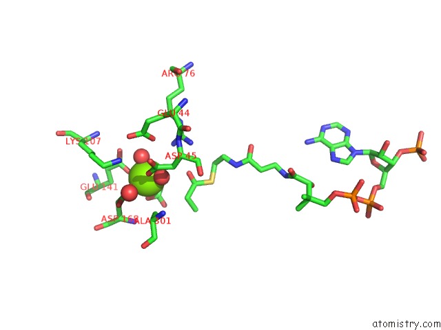
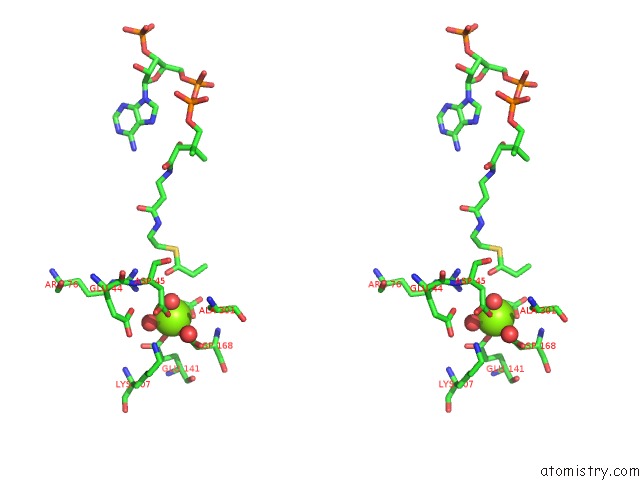
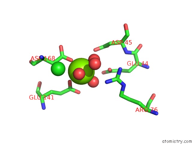
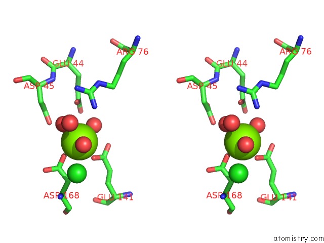
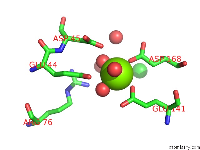
Magnesium binding site 1 out of 6 in 4l9y
Go back to
Magnesium binding site 1 out
of 6 in the Crystal Structure of Rhodobacter Sphaeroides Malyl-Coa Lyase in Complex with Magnesium, Glyoxylate, and Propionyl-Coa
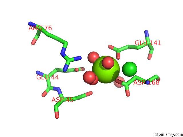
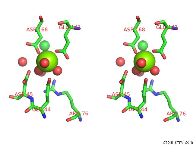
Mono view
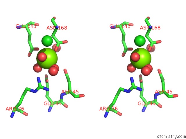
Stereo pair view
Mono view
Stereo pair view
A full contact list of Magnesium with other atoms in the Mg binding
site number 1 of Crystal Structure of Rhodobacter Sphaeroides Malyl-Coa Lyase in Complex with Magnesium, Glyoxylate, and Propionyl-Coa within 5.0Å range:
|
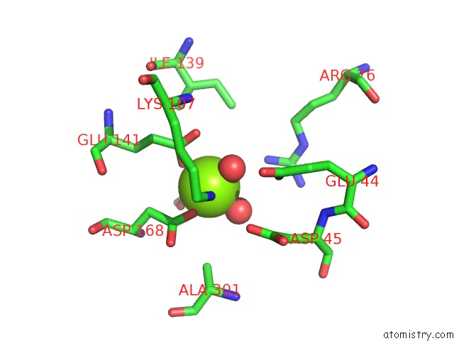
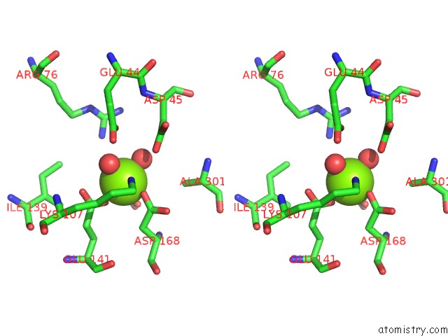
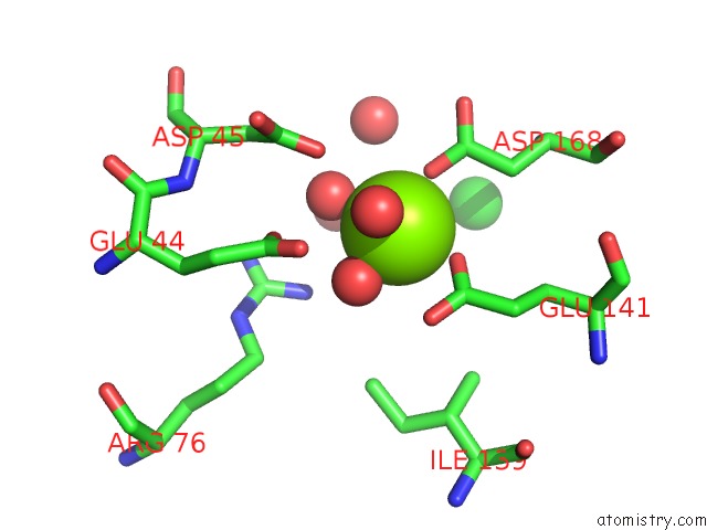
Magnesium binding site 2 out of 6 in 4l9y
Go back to
Magnesium binding site 2 out
of 6 in the Crystal Structure of Rhodobacter Sphaeroides Malyl-Coa Lyase in Complex with Magnesium, Glyoxylate, and Propionyl-Coa
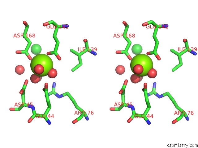
Mono view
Stereo pair view
Mono view
Stereo pair view
A full contact list of Magnesium with other atoms in the Mg binding
site number 2 of Crystal Structure of Rhodobacter Sphaeroides Malyl-Coa Lyase in Complex with Magnesium, Glyoxylate, and Propionyl-Coa within 5.0Å range:
|
Magnesium binding site 3 out of 6 in 4l9y
Go back to
Magnesium binding site 3 out
of 6 in the Crystal Structure of Rhodobacter Sphaeroides Malyl-Coa Lyase in Complex with Magnesium, Glyoxylate, and Propionyl-Coa
Mono view
Stereo pair view
Mono view
Stereo pair view
A full contact list of Magnesium with other atoms in the Mg binding
site number 3 of Crystal Structure of Rhodobacter Sphaeroides Malyl-Coa Lyase in Complex with Magnesium, Glyoxylate, and Propionyl-Coa within 5.0Å range:
|
Magnesium binding site 4 out of 6 in 4l9y
Go back to
Magnesium binding site 4 out
of 6 in the Crystal Structure of Rhodobacter Sphaeroides Malyl-Coa Lyase in Complex with Magnesium, Glyoxylate, and Propionyl-Coa
Mono view
Stereo pair view
Mono view
Stereo pair view
A full contact list of Magnesium with other atoms in the Mg binding
site number 4 of Crystal Structure of Rhodobacter Sphaeroides Malyl-Coa Lyase in Complex with Magnesium, Glyoxylate, and Propionyl-Coa within 5.0Å range:
|
Magnesium binding site 5 out of 6 in 4l9y
Go back to
Magnesium binding site 5 out
of 6 in the Crystal Structure of Rhodobacter Sphaeroides Malyl-Coa Lyase in Complex with Magnesium, Glyoxylate, and Propionyl-Coa
Mono view
Stereo pair view
Mono view
Stereo pair view
A full contact list of Magnesium with other atoms in the Mg binding
site number 5 of Crystal Structure of Rhodobacter Sphaeroides Malyl-Coa Lyase in Complex with Magnesium, Glyoxylate, and Propionyl-Coa within 5.0Å range:
|
Magnesium binding site 6 out of 6 in 4l9y
Go back to
Magnesium binding site 6 out
of 6 in the Crystal Structure of Rhodobacter Sphaeroides Malyl-Coa Lyase in Complex with Magnesium, Glyoxylate, and Propionyl-Coa
Mono view
Stereo pair view
Mono view
Stereo pair view
A full contact list of Magnesium with other atoms in the Mg binding
site number 6 of Crystal Structure of Rhodobacter Sphaeroides Malyl-Coa Lyase in Complex with Magnesium, Glyoxylate, and Propionyl-Coa within 5.0Å range:
|
Reference:
J.Zarzycki,
C.A.Kerfeld.
The Crystal Structures of the Tri-Functional Chloroflexus Aurantiacus and Bi-Functional Rhodobacter Sphaeroides Malyl-Coa Lyases and Comparison with Cite-Like Superfamily Enzymes and Malate Synthases. Bmc Struct.Biol. V. 13 28 2013.
ISSN: ESSN 1472-6807
PubMed: 24206647
DOI: 10.1186/1472-6807-13-28
Page generated: Sat Aug 17 04:29:34 2024
ISSN: ESSN 1472-6807
PubMed: 24206647
DOI: 10.1186/1472-6807-13-28
Last articles
Zn in 9J0NZn in 9J0O
Zn in 9J0P
Zn in 9FJX
Zn in 9EKB
Zn in 9C0F
Zn in 9CAH
Zn in 9CH0
Zn in 9CH3
Zn in 9CH1