Magnesium »
PDB 4la7-4lj9 »
4lem »
Magnesium in PDB 4lem: Crystal Structure of the Delta-Pyrroline-5-Carboxylate Dehydrogenase From Mycobacterium Tuberculosis
Enzymatic activity of Crystal Structure of the Delta-Pyrroline-5-Carboxylate Dehydrogenase From Mycobacterium Tuberculosis
All present enzymatic activity of Crystal Structure of the Delta-Pyrroline-5-Carboxylate Dehydrogenase From Mycobacterium Tuberculosis:
1.5.1.12;
1.5.1.12;
Protein crystallography data
The structure of Crystal Structure of the Delta-Pyrroline-5-Carboxylate Dehydrogenase From Mycobacterium Tuberculosis, PDB code: 4lem
was solved by
T.Lagautriere,
G.Bashiri,
E.N.Baker,
with X-Ray Crystallography technique. A brief refinement statistics is given in the table below:
| Resolution Low / High (Å) | 20.01 / 2.27 |
| Space group | P 31 2 1 |
| Cell size a, b, c (Å), α, β, γ (°) | 164.299, 164.299, 259.114, 90.00, 90.00, 120.00 |
| R / Rfree (%) | 14.8 / 20.4 |
Other elements in 4lem:
The structure of Crystal Structure of the Delta-Pyrroline-5-Carboxylate Dehydrogenase From Mycobacterium Tuberculosis also contains other interesting chemical elements:
| Cobalt | (Co) | 4 atoms |
Magnesium Binding Sites:
The binding sites of Magnesium atom in the Crystal Structure of the Delta-Pyrroline-5-Carboxylate Dehydrogenase From Mycobacterium Tuberculosis
(pdb code 4lem). This binding sites where shown within
5.0 Angstroms radius around Magnesium atom.
In total 3 binding sites of Magnesium where determined in the Crystal Structure of the Delta-Pyrroline-5-Carboxylate Dehydrogenase From Mycobacterium Tuberculosis, PDB code: 4lem:
Jump to Magnesium binding site number: 1; 2; 3;
In total 3 binding sites of Magnesium where determined in the Crystal Structure of the Delta-Pyrroline-5-Carboxylate Dehydrogenase From Mycobacterium Tuberculosis, PDB code: 4lem:
Jump to Magnesium binding site number: 1; 2; 3;
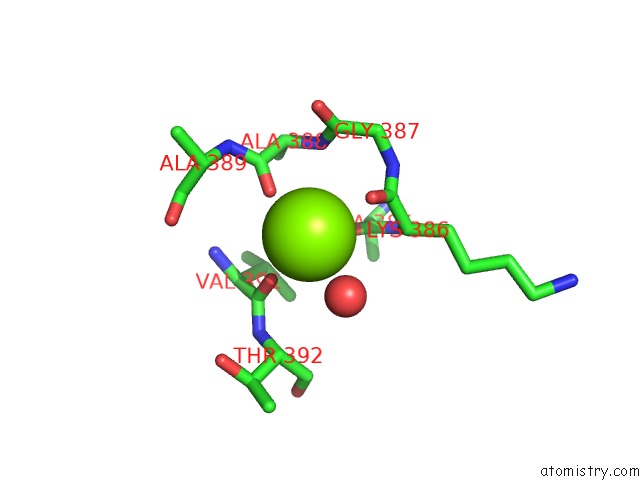
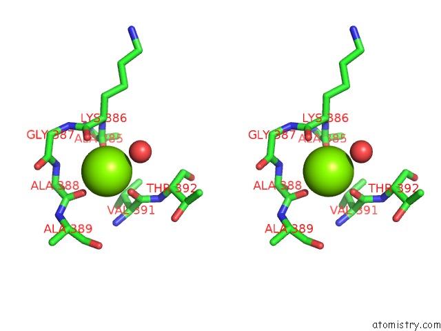
Magnesium binding site 1 out of 3 in 4lem
Go back to
Magnesium binding site 1 out
of 3 in the Crystal Structure of the Delta-Pyrroline-5-Carboxylate Dehydrogenase From Mycobacterium Tuberculosis
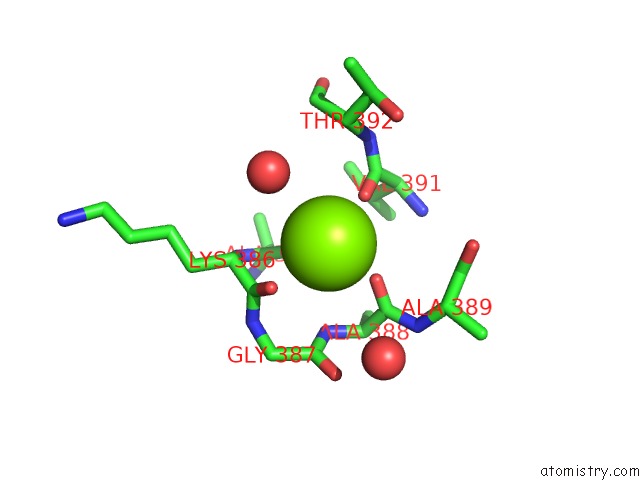
Mono view
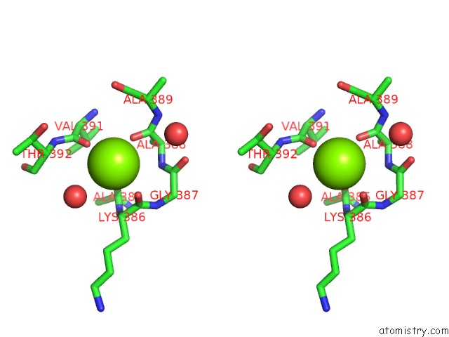
Stereo pair view
Mono view
Stereo pair view
A full contact list of Magnesium with other atoms in the Mg binding
site number 1 of Crystal Structure of the Delta-Pyrroline-5-Carboxylate Dehydrogenase From Mycobacterium Tuberculosis within 5.0Å range:
|
Magnesium binding site 2 out of 3 in 4lem
Go back to
Magnesium binding site 2 out
of 3 in the Crystal Structure of the Delta-Pyrroline-5-Carboxylate Dehydrogenase From Mycobacterium Tuberculosis
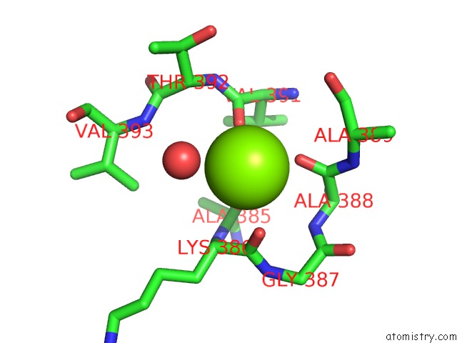
Mono view
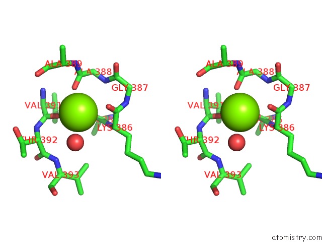
Stereo pair view
Mono view
Stereo pair view
A full contact list of Magnesium with other atoms in the Mg binding
site number 2 of Crystal Structure of the Delta-Pyrroline-5-Carboxylate Dehydrogenase From Mycobacterium Tuberculosis within 5.0Å range:
|
Magnesium binding site 3 out of 3 in 4lem
Go back to
Magnesium binding site 3 out
of 3 in the Crystal Structure of the Delta-Pyrroline-5-Carboxylate Dehydrogenase From Mycobacterium Tuberculosis
Mono view
Stereo pair view
Mono view
Stereo pair view
A full contact list of Magnesium with other atoms in the Mg binding
site number 3 of Crystal Structure of the Delta-Pyrroline-5-Carboxylate Dehydrogenase From Mycobacterium Tuberculosis within 5.0Å range:
|
Reference:
T.Lagautriere,
G.Bashiri,
E.N.Baker.
Crystal Structure of the Delta-Pyrroline-5-Carboxylate Dehydrogenase From Mycobacterium Tuberculosis To Be Published.
Page generated: Mon Aug 11 18:31:42 2025
Last articles
Mg in 5DH9Mg in 5DH4
Mg in 5DGH
Mg in 5DGT
Mg in 5DGK
Mg in 5DEI
Mg in 5DGI
Mg in 5DGD
Mg in 5DGB
Mg in 5DGA