Magnesium »
PDB 4lj9-4lse »
4lnj »
Magnesium in PDB 4lnj: Structure of Escherichia Coli Threonine Aldolase in Unliganded Form
Enzymatic activity of Structure of Escherichia Coli Threonine Aldolase in Unliganded Form
All present enzymatic activity of Structure of Escherichia Coli Threonine Aldolase in Unliganded Form:
4.1.2.5;
4.1.2.5;
Protein crystallography data
The structure of Structure of Escherichia Coli Threonine Aldolase in Unliganded Form, PDB code: 4lnj
was solved by
M.K.Safo,
R.Contestabile,
S.G.Remesh,
with X-Ray Crystallography technique. A brief refinement statistics is given in the table below:
| Resolution Low / High (Å) | 29.35 / 2.10 |
| Space group | C 2 2 21 |
| Cell size a, b, c (Å), α, β, γ (°) | 76.620, 100.790, 176.080, 90.00, 90.00, 90.00 |
| R / Rfree (%) | 20.9 / 27.9 |
Magnesium Binding Sites:
The binding sites of Magnesium atom in the Structure of Escherichia Coli Threonine Aldolase in Unliganded Form
(pdb code 4lnj). This binding sites where shown within
5.0 Angstroms radius around Magnesium atom.
In total 3 binding sites of Magnesium where determined in the Structure of Escherichia Coli Threonine Aldolase in Unliganded Form, PDB code: 4lnj:
Jump to Magnesium binding site number: 1; 2; 3;
In total 3 binding sites of Magnesium where determined in the Structure of Escherichia Coli Threonine Aldolase in Unliganded Form, PDB code: 4lnj:
Jump to Magnesium binding site number: 1; 2; 3;
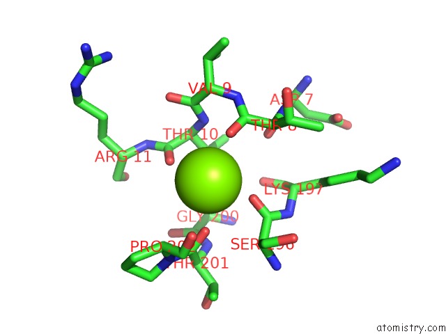
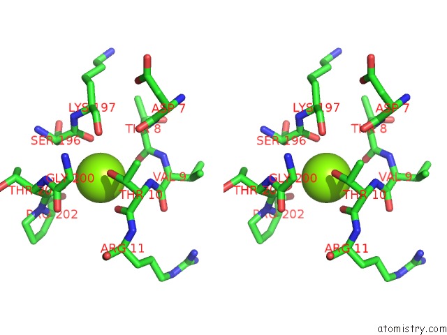
Magnesium binding site 1 out of 3 in 4lnj
Go back to
Magnesium binding site 1 out
of 3 in the Structure of Escherichia Coli Threonine Aldolase in Unliganded Form
Mono view
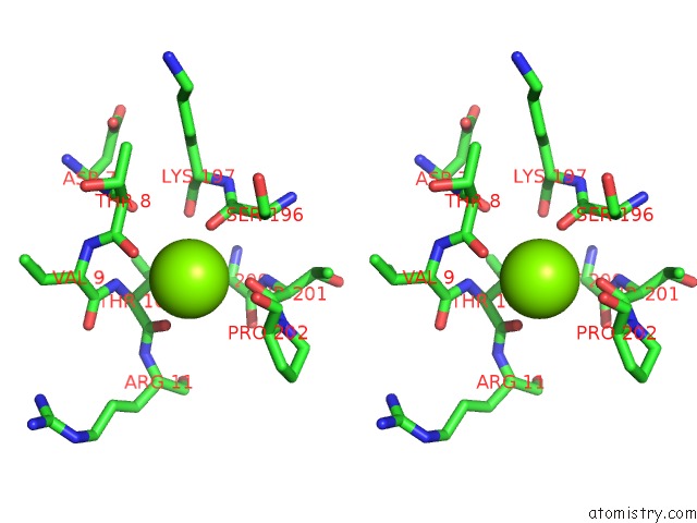
Stereo pair view
Mono view
Stereo pair view
A full contact list of Magnesium with other atoms in the Mg binding
site number 1 of Structure of Escherichia Coli Threonine Aldolase in Unliganded Form within 5.0Å range:
|
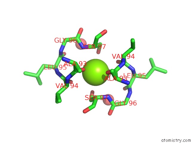
Magnesium binding site 2 out of 3 in 4lnj
Go back to
Magnesium binding site 2 out
of 3 in the Structure of Escherichia Coli Threonine Aldolase in Unliganded Form
Mono view

Stereo pair view
Mono view
Stereo pair view
A full contact list of Magnesium with other atoms in the Mg binding
site number 2 of Structure of Escherichia Coli Threonine Aldolase in Unliganded Form within 5.0Å range:
|
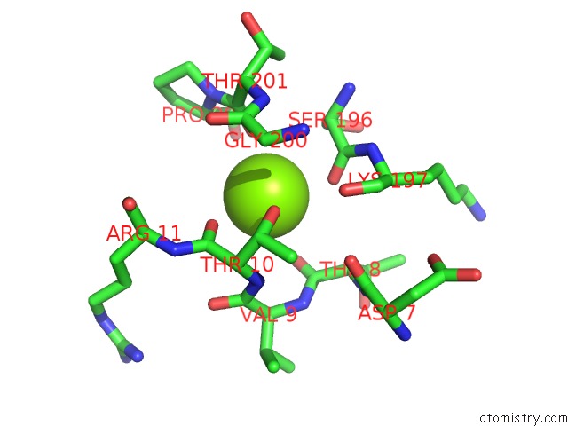
Magnesium binding site 3 out of 3 in 4lnj
Go back to
Magnesium binding site 3 out
of 3 in the Structure of Escherichia Coli Threonine Aldolase in Unliganded Form
Mono view
Stereo pair view
Mono view
Stereo pair view
A full contact list of Magnesium with other atoms in the Mg binding
site number 3 of Structure of Escherichia Coli Threonine Aldolase in Unliganded Form within 5.0Å range:
|
Reference:
M.L.Di Salvo,
S.G.Remesh,
M.Vivoli,
M.S.Ghatge,
A.Paiardini,
S.D'aguanno,
M.K.Safo,
R.Contestabile.
On the Catalytic Mechanism and Stereospecificity of Escherichia Coli L-Threonine Aldolase. Febs J. V. 281 129 2014.
ISSN: ISSN 1742-464X
PubMed: 24165453
DOI: 10.1111/FEBS.12581
Page generated: Mon Aug 19 19:49:16 2024
ISSN: ISSN 1742-464X
PubMed: 24165453
DOI: 10.1111/FEBS.12581
Last articles
Zn in 9JYWZn in 9IR4
Zn in 9IR3
Zn in 9GMX
Zn in 9GMW
Zn in 9JEJ
Zn in 9ERF
Zn in 9ERE
Zn in 9EGV
Zn in 9EGW