Magnesium »
PDB 4m3n-4mfe »
4mar »
Magnesium in PDB 4mar: Crystal Structure of Purine Nucleoside Phosphorylase From Meiothermus Ruber Dsm 1279 Complexed with Sulfate.
Enzymatic activity of Crystal Structure of Purine Nucleoside Phosphorylase From Meiothermus Ruber Dsm 1279 Complexed with Sulfate.
All present enzymatic activity of Crystal Structure of Purine Nucleoside Phosphorylase From Meiothermus Ruber Dsm 1279 Complexed with Sulfate.:
2.4.2.1;
2.4.2.1;
Protein crystallography data
The structure of Crystal Structure of Purine Nucleoside Phosphorylase From Meiothermus Ruber Dsm 1279 Complexed with Sulfate., PDB code: 4mar
was solved by
V.N.Malashkevich,
R.Bhosle,
R.Toro,
B.Hillerich,
A.Gizzi,
S.Garforth,
A.Kar,
M.K.Chan,
J.Lafluer,
H.Patel,
B.Matikainen,
S.Chamala,
S.Lim,
A.Celikgil,
G.Villegas,
B.Evans,
J.Love,
A.Fiser,
K.Khafizov,
R.Seidel,
J.B.Bonanno,
S.C.Almo,
New York Structural Genomics Researchconsortium (Nysgrc),
with X-Ray Crystallography technique. A brief refinement statistics is given in the table below:
| Resolution Low / High (Å) | 32.56 / 2.16 |
| Space group | C 2 2 21 |
| Cell size a, b, c (Å), α, β, γ (°) | 57.403, 189.103, 152.235, 90.00, 90.00, 90.00 |
| R / Rfree (%) | 18.8 / 22.6 |
Magnesium Binding Sites:
The binding sites of Magnesium atom in the Crystal Structure of Purine Nucleoside Phosphorylase From Meiothermus Ruber Dsm 1279 Complexed with Sulfate.
(pdb code 4mar). This binding sites where shown within
5.0 Angstroms radius around Magnesium atom.
In total only one binding site of Magnesium was determined in the Crystal Structure of Purine Nucleoside Phosphorylase From Meiothermus Ruber Dsm 1279 Complexed with Sulfate., PDB code: 4mar:
In total only one binding site of Magnesium was determined in the Crystal Structure of Purine Nucleoside Phosphorylase From Meiothermus Ruber Dsm 1279 Complexed with Sulfate., PDB code: 4mar:
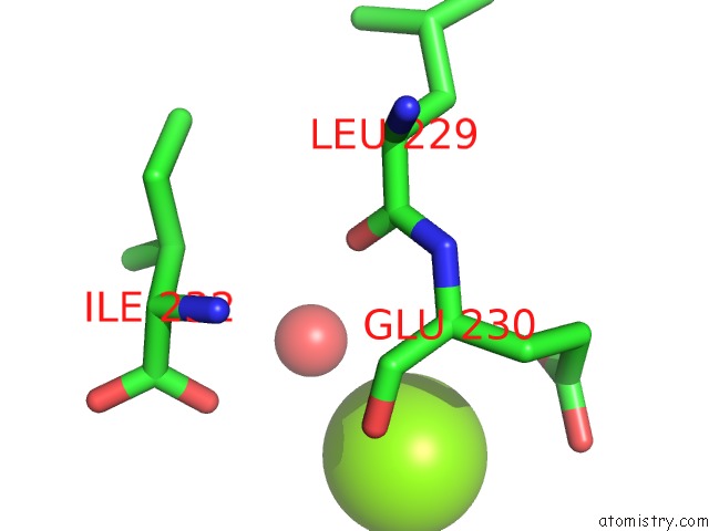
Magnesium binding site 1 out of 1 in 4mar
Go back to
Magnesium binding site 1 out
of 1 in the Crystal Structure of Purine Nucleoside Phosphorylase From Meiothermus Ruber Dsm 1279 Complexed with Sulfate.
Mono view

Stereo pair view
Mono view
Stereo pair view
A full contact list of Magnesium with other atoms in the Mg binding
site number 1 of Crystal Structure of Purine Nucleoside Phosphorylase From Meiothermus Ruber Dsm 1279 Complexed with Sulfate. within 5.0Å range:
|
Reference:
V.N.Malashkevich,
R.Bhosle,
R.Toro,
B.Hillerich,
A.Gizzi,
S.Garforth,
A.Kar,
M.K.Chan,
J.Lafluer,
H.Patel,
B.Matikainen,
S.Chamala,
S.Lim,
A.Celikgil,
G.Villegas,
B.Evans,
J.Love,
A.Fiser,
K.Khafizov,
R.Seidel,
J.B.Bonanno,
S.C.Almo.
Crystal Structure of Purine Nucleoside Phosphorylase From Meiothermus Ruber Dsm 1279 Complexed with Sulfate. To Be Published.
Page generated: Mon Aug 11 20:21:16 2025
Last articles
Mg in 4UREMg in 4UQP
Mg in 4UQL
Mg in 4UQO
Mg in 4UOO
Mg in 4UPH
Mg in 4UPV
Mg in 4UOP
Mg in 4UOG
Mg in 4UOH