Magnesium »
PDB 4m3n-4mfe »
4mfd »
Magnesium in PDB 4mfd: Structure of the Carboxyl Transferase Domain From Rhizobium Etli Pyruvate Carboxylase with Oxalate
Enzymatic activity of Structure of the Carboxyl Transferase Domain From Rhizobium Etli Pyruvate Carboxylase with Oxalate
All present enzymatic activity of Structure of the Carboxyl Transferase Domain From Rhizobium Etli Pyruvate Carboxylase with Oxalate:
6.4.1.1;
6.4.1.1;
Protein crystallography data
The structure of Structure of the Carboxyl Transferase Domain From Rhizobium Etli Pyruvate Carboxylase with Oxalate, PDB code: 4mfd
was solved by
A.D.Lietzan,
M.St. Maurice,
with X-Ray Crystallography technique. A brief refinement statistics is given in the table below:
| Resolution Low / High (Å) | 48.26 / 2.55 |
| Space group | P 21 21 21 |
| Cell size a, b, c (Å), α, β, γ (°) | 85.657, 157.368, 244.827, 90.00, 90.00, 90.00 |
| R / Rfree (%) | 19.3 / 24 |
Other elements in 4mfd:
The structure of Structure of the Carboxyl Transferase Domain From Rhizobium Etli Pyruvate Carboxylase with Oxalate also contains other interesting chemical elements:
| Chlorine | (Cl) | 4 atoms |
| Zinc | (Zn) | 4 atoms |
Magnesium Binding Sites:
The binding sites of Magnesium atom in the Structure of the Carboxyl Transferase Domain From Rhizobium Etli Pyruvate Carboxylase with Oxalate
(pdb code 4mfd). This binding sites where shown within
5.0 Angstroms radius around Magnesium atom.
In total 4 binding sites of Magnesium where determined in the Structure of the Carboxyl Transferase Domain From Rhizobium Etli Pyruvate Carboxylase with Oxalate, PDB code: 4mfd:
Jump to Magnesium binding site number: 1; 2; 3; 4;
In total 4 binding sites of Magnesium where determined in the Structure of the Carboxyl Transferase Domain From Rhizobium Etli Pyruvate Carboxylase with Oxalate, PDB code: 4mfd:
Jump to Magnesium binding site number: 1; 2; 3; 4;
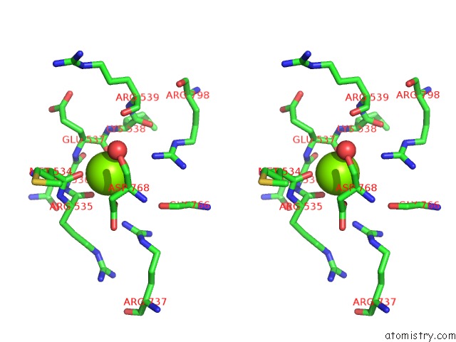
Magnesium binding site 1 out of 4 in 4mfd
Go back to
Magnesium binding site 1 out
of 4 in the Structure of the Carboxyl Transferase Domain From Rhizobium Etli Pyruvate Carboxylase with Oxalate
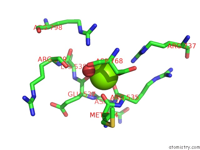
Mono view
Stereo pair view
Mono view
Stereo pair view
A full contact list of Magnesium with other atoms in the Mg binding
site number 1 of Structure of the Carboxyl Transferase Domain From Rhizobium Etli Pyruvate Carboxylase with Oxalate within 5.0Å range:
|
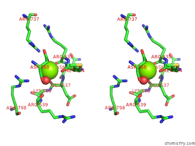
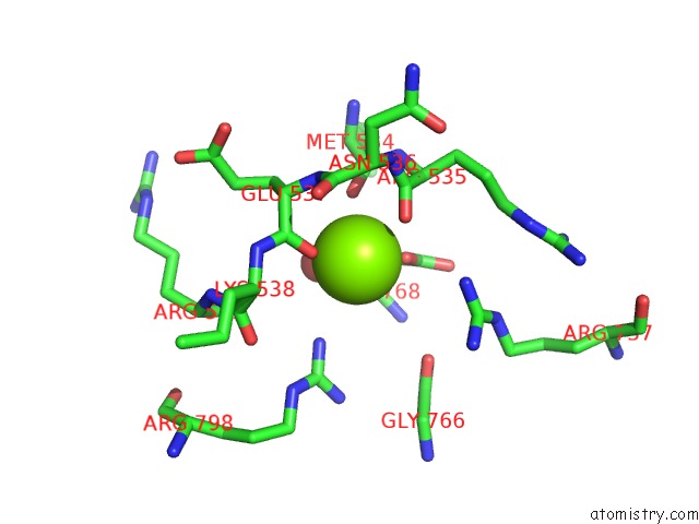
Magnesium binding site 2 out of 4 in 4mfd
Go back to
Magnesium binding site 2 out
of 4 in the Structure of the Carboxyl Transferase Domain From Rhizobium Etli Pyruvate Carboxylase with Oxalate
Mono view
Stereo pair view
Mono view
Stereo pair view
A full contact list of Magnesium with other atoms in the Mg binding
site number 2 of Structure of the Carboxyl Transferase Domain From Rhizobium Etli Pyruvate Carboxylase with Oxalate within 5.0Å range:
|
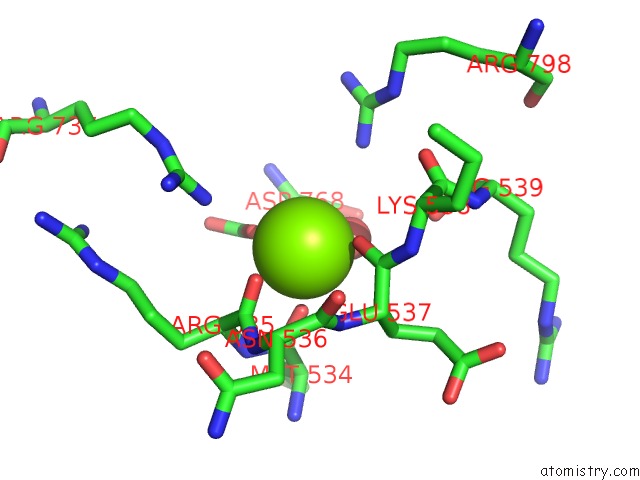
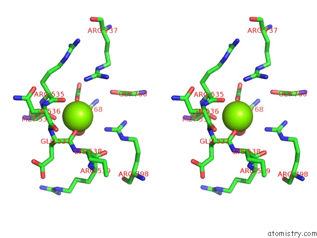
Magnesium binding site 3 out of 4 in 4mfd
Go back to
Magnesium binding site 3 out
of 4 in the Structure of the Carboxyl Transferase Domain From Rhizobium Etli Pyruvate Carboxylase with Oxalate

Mono view
Stereo pair view
Mono view
Stereo pair view
A full contact list of Magnesium with other atoms in the Mg binding
site number 3 of Structure of the Carboxyl Transferase Domain From Rhizobium Etli Pyruvate Carboxylase with Oxalate within 5.0Å range:
|
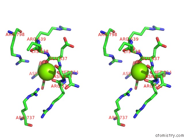
Magnesium binding site 4 out of 4 in 4mfd
Go back to
Magnesium binding site 4 out
of 4 in the Structure of the Carboxyl Transferase Domain From Rhizobium Etli Pyruvate Carboxylase with Oxalate
Mono view
Stereo pair view
Mono view
Stereo pair view
A full contact list of Magnesium with other atoms in the Mg binding
site number 4 of Structure of the Carboxyl Transferase Domain From Rhizobium Etli Pyruvate Carboxylase with Oxalate within 5.0Å range:
|
Reference:
A.D.Lietzan,
M.St. Maurice.
Insights Into the Carboxyltransferase Reaction of Pyruvate Carboxylase From the Structures of Bound Product and Intermediate Analogs. Biochem.Biophys.Res.Commun. V. 441 377 2013.
ISSN: ISSN 0006-291X
PubMed: 24157795
DOI: 10.1016/J.BBRC.2013.10.066
Page generated: Mon Aug 19 20:22:19 2024
ISSN: ISSN 0006-291X
PubMed: 24157795
DOI: 10.1016/J.BBRC.2013.10.066
Last articles
Ca in 5T50Ca in 5T4Z
Ca in 5T0X
Ca in 5T3H
Ca in 5T2N
Ca in 5T2O
Ca in 5T2H
Ca in 5SZQ
Ca in 5SZO
Ca in 5SZP