Magnesium »
PDB 4mfe-4mpj »
4mo7 »
Magnesium in PDB 4mo7: Crystal Structure of Superantigen Pfit
Protein crystallography data
The structure of Crystal Structure of Superantigen Pfit, PDB code: 4mo7
was solved by
L.H.Liu,
H.Chen,
H.M.Li,
with X-Ray Crystallography technique. A brief refinement statistics is given in the table below:
| Resolution Low / High (Å) | 25.19 / 1.70 |
| Space group | C 1 2 1 |
| Cell size a, b, c (Å), α, β, γ (°) | 37.226, 93.661, 53.557, 90.00, 109.82, 90.00 |
| R / Rfree (%) | 18.9 / 23.4 |
Magnesium Binding Sites:
The binding sites of Magnesium atom in the Crystal Structure of Superantigen Pfit
(pdb code 4mo7). This binding sites where shown within
5.0 Angstroms radius around Magnesium atom.
In total only one binding site of Magnesium was determined in the Crystal Structure of Superantigen Pfit, PDB code: 4mo7:
In total only one binding site of Magnesium was determined in the Crystal Structure of Superantigen Pfit, PDB code: 4mo7:
Magnesium binding site 1 out of 1 in 4mo7
Go back to
Magnesium binding site 1 out
of 1 in the Crystal Structure of Superantigen Pfit

Mono view
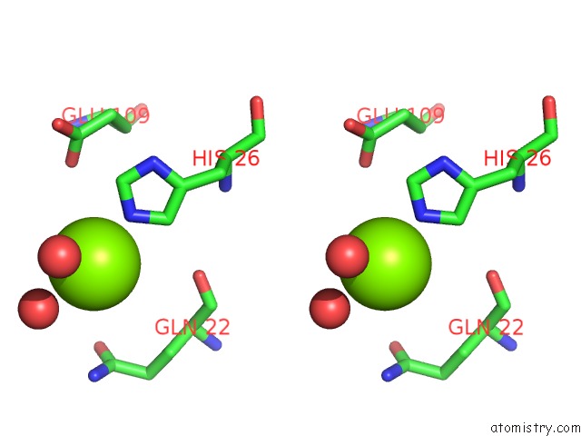
Stereo pair view
Mono view
Stereo pair view
A full contact list of Magnesium with other atoms in the Mg binding
site number 1 of Crystal Structure of Superantigen Pfit within 5.0Å range:
|
Reference:
L.Liu,
H.Chen,
M.B.Brecher,
Z.Li,
B.Wei,
B.Nandi,
J.Zhang,
H.Ling,
G.Winslow,
J.Braun,
H.Li.
Pfit Is A Structurally Novel Crohn'S Disease-Associated Superantigen. Plos Pathog. V. 9 03837 2013.
ISSN: ISSN 1553-7366
PubMed: 24385909
DOI: 10.1371/JOURNAL.PPAT.1003837
Page generated: Mon Aug 19 23:17:06 2024
ISSN: ISSN 1553-7366
PubMed: 24385909
DOI: 10.1371/JOURNAL.PPAT.1003837
Last articles
Zn in 9J0NZn in 9J0O
Zn in 9J0P
Zn in 9FJX
Zn in 9EKB
Zn in 9C0F
Zn in 9CAH
Zn in 9CH0
Zn in 9CH3
Zn in 9CH1