Magnesium »
PDB 4mpo-4naa »
4mum »
Magnesium in PDB 4mum: Crystal Structure of Mitochondrial 5'(3')-Deoxy Ribonucleotidase Alternative Spliced Variant
Protein crystallography data
The structure of Crystal Structure of Mitochondrial 5'(3')-Deoxy Ribonucleotidase Alternative Spliced Variant, PDB code: 4mum
was solved by
P.Pachl,
P.Rezacova,
with X-Ray Crystallography technique. A brief refinement statistics is given in the table below:
| Resolution Low / High (Å) | 46.66 / 1.27 |
| Space group | P 43 21 2 |
| Cell size a, b, c (Å), α, β, γ (°) | 73.372, 73.372, 106.672, 90.00, 90.00, 90.00 |
| R / Rfree (%) | 13.1 / 14.9 |
Other elements in 4mum:
The structure of Crystal Structure of Mitochondrial 5'(3')-Deoxy Ribonucleotidase Alternative Spliced Variant also contains other interesting chemical elements:
| Chlorine | (Cl) | 1 atom |
Magnesium Binding Sites:
The binding sites of Magnesium atom in the Crystal Structure of Mitochondrial 5'(3')-Deoxy Ribonucleotidase Alternative Spliced Variant
(pdb code 4mum). This binding sites where shown within
5.0 Angstroms radius around Magnesium atom.
In total only one binding site of Magnesium was determined in the Crystal Structure of Mitochondrial 5'(3')-Deoxy Ribonucleotidase Alternative Spliced Variant, PDB code: 4mum:
In total only one binding site of Magnesium was determined in the Crystal Structure of Mitochondrial 5'(3')-Deoxy Ribonucleotidase Alternative Spliced Variant, PDB code: 4mum:
Magnesium binding site 1 out of 1 in 4mum
Go back to
Magnesium binding site 1 out
of 1 in the Crystal Structure of Mitochondrial 5'(3')-Deoxy Ribonucleotidase Alternative Spliced Variant
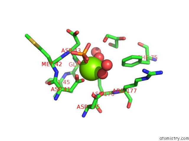
Mono view
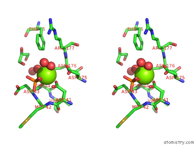
Stereo pair view
Mono view
Stereo pair view
A full contact list of Magnesium with other atoms in the Mg binding
site number 1 of Crystal Structure of Mitochondrial 5'(3')-Deoxy Ribonucleotidase Alternative Spliced Variant within 5.0Å range:
|
Reference:
P.Pachl,
M.Fabry,
V.Veverka,
J.Brynda,
P.Rezacova.
Kinetic and Structural Characterization of An Alternatively Spliced Variant of Human Mitochondrial 5'(3')-Deoxyribonucleotidase. J Enzyme Inhib Med Chem 2014.
ISSN: ESSN 1475-6374
PubMed: 24506201
DOI: 10.3109/14756366.2013.879577
Page generated: Mon Aug 19 23:23:21 2024
ISSN: ESSN 1475-6374
PubMed: 24506201
DOI: 10.3109/14756366.2013.879577
Last articles
Zn in 9J0NZn in 9J0O
Zn in 9J0P
Zn in 9FJX
Zn in 9EKB
Zn in 9C0F
Zn in 9CAH
Zn in 9CH0
Zn in 9CH3
Zn in 9CH1