Magnesium »
PDB 4nbm-4nlz »
4ni5 »
Magnesium in PDB 4ni5: Crystal Structure of A Short Chain Dehydrogenase From Brucella Suis
Protein crystallography data
The structure of Crystal Structure of A Short Chain Dehydrogenase From Brucella Suis, PDB code: 4ni5
was solved by
D.M.Dranow,
J.Abendroth,
T.E.Edwards,
D.Lorimer,
Seattle Structuralgenomics Center For Infectious Disease (Ssgcid),
with X-Ray Crystallography technique. A brief refinement statistics is given in the table below:
| Resolution Low / High (Å) | 50.00 / 1.70 |
| Space group | C 2 2 21 |
| Cell size a, b, c (Å), α, β, γ (°) | 62.440, 130.390, 114.120, 90.00, 90.00, 90.00 |
| R / Rfree (%) | 15.1 / 18.1 |
Magnesium Binding Sites:
The binding sites of Magnesium atom in the Crystal Structure of A Short Chain Dehydrogenase From Brucella Suis
(pdb code 4ni5). This binding sites where shown within
5.0 Angstroms radius around Magnesium atom.
In total only one binding site of Magnesium was determined in the Crystal Structure of A Short Chain Dehydrogenase From Brucella Suis, PDB code: 4ni5:
In total only one binding site of Magnesium was determined in the Crystal Structure of A Short Chain Dehydrogenase From Brucella Suis, PDB code: 4ni5:
Magnesium binding site 1 out of 1 in 4ni5
Go back to
Magnesium binding site 1 out
of 1 in the Crystal Structure of A Short Chain Dehydrogenase From Brucella Suis
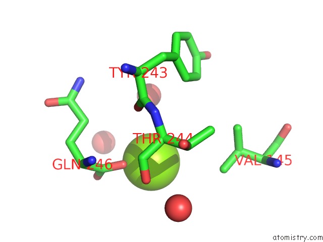
Mono view
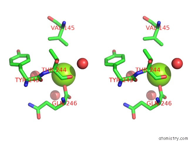
Stereo pair view
Mono view
Stereo pair view
A full contact list of Magnesium with other atoms in the Mg binding
site number 1 of Crystal Structure of A Short Chain Dehydrogenase From Brucella Suis within 5.0Å range:
|
Reference:
D.M.Dranow,
J.Abendroth,
T.E.Edwards,
D.Lorimer.
Crystal Structure of A Short Chain Dehydrogenase From Brucella Suis To Be Published.
Page generated: Mon Aug 11 20:43:39 2025
Last articles
Mg in 4V26Mg in 4V2G
Mg in 4V1T
Mg in 4V25
Mg in 4V1V
Mg in 4V1O
Mg in 4V1N
Mg in 4V1M
Mg in 4V0R
Mg in 4V0O