Magnesium »
PDB 4nlz-4nxm »
4nob »
Magnesium in PDB 4nob: Crystal Structure of the 1ST Ig Domain From Mouse Polymeric Immunoglobulin Receptor [Psi-Nysgrc-006220]
Protein crystallography data
The structure of Crystal Structure of the 1ST Ig Domain From Mouse Polymeric Immunoglobulin Receptor [Psi-Nysgrc-006220], PDB code: 4nob
was solved by
P.R.Kumar,
R.Banu,
R.Bhosle,
D.A.Calarese,
A.Celikgil,
S.Chamala,
M.K.Chan,
S.Chowdhury,
A.Fiser,
S.J.Garforth,
A.S.Glenn,
B.Hillerich,
K.Khafizov,
J.Attonito,
J.D.Love,
H.Patel,
R.Patel,
R.D.Seidel,
B.Smith,
M.Stead,
A.Casadevall,
S.C.Almo,
New York Structural Genomics Researchconsortium (Nysgrc),
Atoms-To-Animals: The Immune Function Network(Ifn),
with X-Ray Crystallography technique. A brief refinement statistics is given in the table below:
| Resolution Low / High (Å) | 37.37 / 1.51 |
| Space group | P 21 21 21 |
| Cell size a, b, c (Å), α, β, γ (°) | 40.275, 46.803, 62.062, 90.00, 90.00, 90.00 |
| R / Rfree (%) | 16.1 / 18.9 |
Magnesium Binding Sites:
The binding sites of Magnesium atom in the Crystal Structure of the 1ST Ig Domain From Mouse Polymeric Immunoglobulin Receptor [Psi-Nysgrc-006220]
(pdb code 4nob). This binding sites where shown within
5.0 Angstroms radius around Magnesium atom.
In total only one binding site of Magnesium was determined in the Crystal Structure of the 1ST Ig Domain From Mouse Polymeric Immunoglobulin Receptor [Psi-Nysgrc-006220], PDB code: 4nob:
In total only one binding site of Magnesium was determined in the Crystal Structure of the 1ST Ig Domain From Mouse Polymeric Immunoglobulin Receptor [Psi-Nysgrc-006220], PDB code: 4nob:
Magnesium binding site 1 out of 1 in 4nob
Go back to
Magnesium binding site 1 out
of 1 in the Crystal Structure of the 1ST Ig Domain From Mouse Polymeric Immunoglobulin Receptor [Psi-Nysgrc-006220]
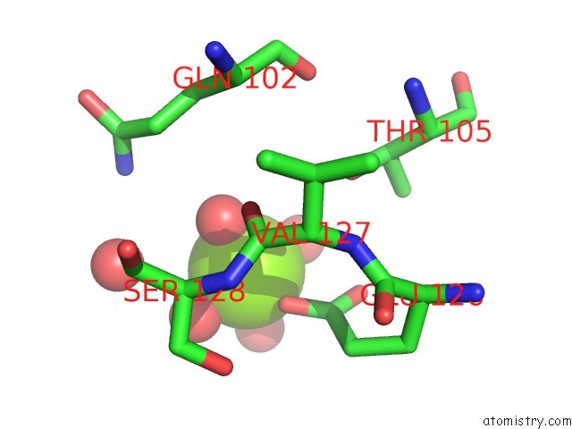
Mono view
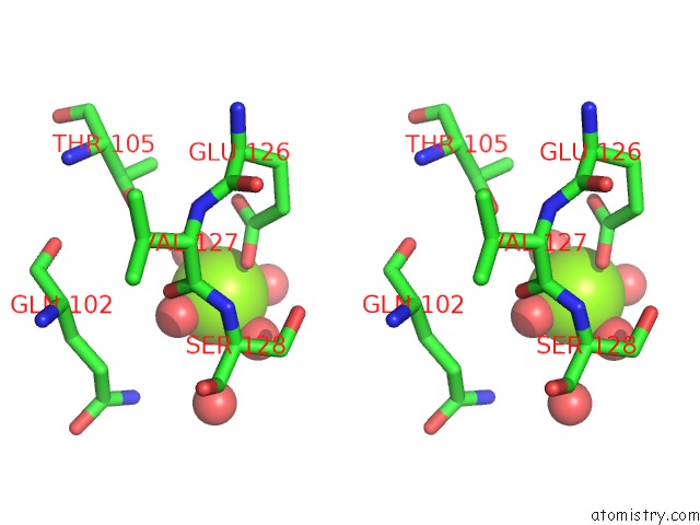
Stereo pair view
Mono view
Stereo pair view
A full contact list of Magnesium with other atoms in the Mg binding
site number 1 of Crystal Structure of the 1ST Ig Domain From Mouse Polymeric Immunoglobulin Receptor [Psi-Nysgrc-006220] within 5.0Å range:
|
Reference:
P.R.Kumar,
A.Casadevall,
S.C.Almo.
Crystal Structure of the 1ST Ig Domain From Mouse Polymeric Immunoglobulin Receptor To Be Published.
Page generated: Mon Aug 19 23:43:30 2024
Last articles
Zn in 9J0NZn in 9J0O
Zn in 9J0P
Zn in 9FJX
Zn in 9EKB
Zn in 9C0F
Zn in 9CAH
Zn in 9CH0
Zn in 9CH3
Zn in 9CH1