Magnesium »
PDB 4nlz-4nxm »
4nq3 »
Magnesium in PDB 4nq3: Crystal Structure of Cyanuic Acid Hydrolase From A. Caulinodans
Protein crystallography data
The structure of Crystal Structure of Cyanuic Acid Hydrolase From A. Caulinodans, PDB code: 4nq3
was solved by
S.Cho,
K.Shi,
H.Aihara,
with X-Ray Crystallography technique. A brief refinement statistics is given in the table below:
| Resolution Low / High (Å) | 40.79 / 2.70 |
| Space group | I 41 2 2 |
| Cell size a, b, c (Å), α, β, γ (°) | 237.866, 237.866, 105.313, 90.00, 90.00, 90.00 |
| R / Rfree (%) | 15.9 / 19.1 |
Magnesium Binding Sites:
The binding sites of Magnesium atom in the Crystal Structure of Cyanuic Acid Hydrolase From A. Caulinodans
(pdb code 4nq3). This binding sites where shown within
5.0 Angstroms radius around Magnesium atom.
In total 3 binding sites of Magnesium where determined in the Crystal Structure of Cyanuic Acid Hydrolase From A. Caulinodans, PDB code: 4nq3:
Jump to Magnesium binding site number: 1; 2; 3;
In total 3 binding sites of Magnesium where determined in the Crystal Structure of Cyanuic Acid Hydrolase From A. Caulinodans, PDB code: 4nq3:
Jump to Magnesium binding site number: 1; 2; 3;
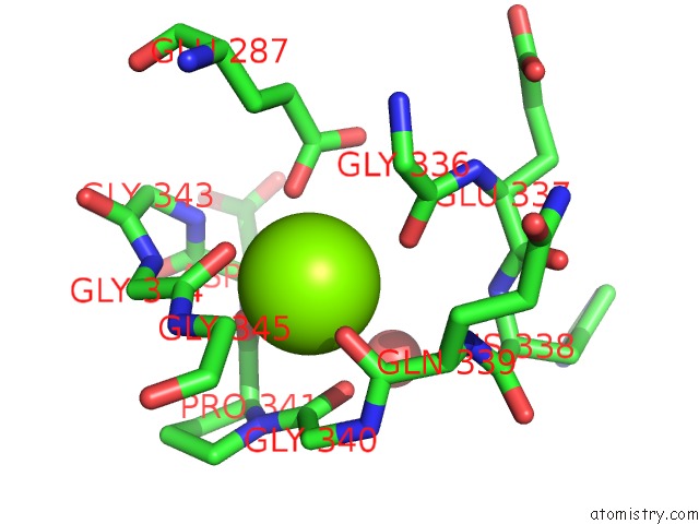
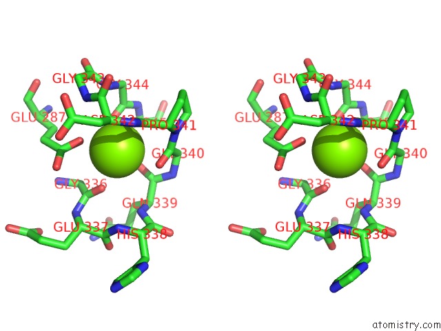
Magnesium binding site 1 out of 3 in 4nq3
Go back to
Magnesium binding site 1 out
of 3 in the Crystal Structure of Cyanuic Acid Hydrolase From A. Caulinodans
Mono view

Stereo pair view
Mono view
Stereo pair view
A full contact list of Magnesium with other atoms in the Mg binding
site number 1 of Crystal Structure of Cyanuic Acid Hydrolase From A. Caulinodans within 5.0Å range:
|
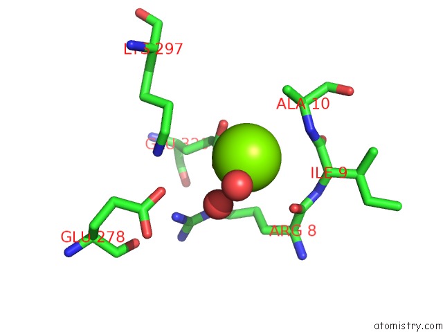
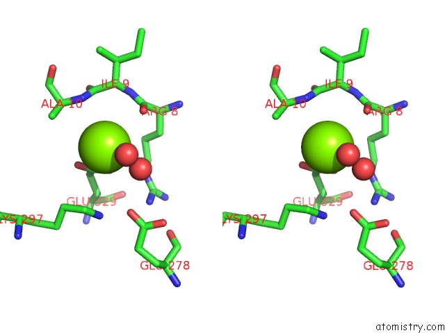
Magnesium binding site 2 out of 3 in 4nq3
Go back to
Magnesium binding site 2 out
of 3 in the Crystal Structure of Cyanuic Acid Hydrolase From A. Caulinodans
Mono view
Stereo pair view
Mono view
Stereo pair view
A full contact list of Magnesium with other atoms in the Mg binding
site number 2 of Crystal Structure of Cyanuic Acid Hydrolase From A. Caulinodans within 5.0Å range:
|
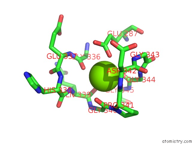
Magnesium binding site 3 out of 3 in 4nq3
Go back to
Magnesium binding site 3 out
of 3 in the Crystal Structure of Cyanuic Acid Hydrolase From A. Caulinodans
Mono view
Stereo pair view
Mono view
Stereo pair view
A full contact list of Magnesium with other atoms in the Mg binding
site number 3 of Crystal Structure of Cyanuic Acid Hydrolase From A. Caulinodans within 5.0Å range:
|
Reference:
S.Cho,
K.Shi,
J.L.Seffernick,
A.G.Dodge,
L.P.Wackett,
H.Aihara.
Cyanuric Acid Hydrolase From Azorhizobium Caulinodans Ors 571: Crystal Structure and Insights Into A New Class of Ser-Lys Dyad Proteins. Plos One V. 9 99349 2014.
ISSN: ESSN 1932-6203
PubMed: 24915109
DOI: 10.1371/JOURNAL.PONE.0099349
Page generated: Mon Aug 19 23:44:44 2024
ISSN: ESSN 1932-6203
PubMed: 24915109
DOI: 10.1371/JOURNAL.PONE.0099349
Last articles
Zn in 9JYWZn in 9IR4
Zn in 9IR3
Zn in 9GMX
Zn in 9GMW
Zn in 9JEJ
Zn in 9ERF
Zn in 9ERE
Zn in 9EGV
Zn in 9EGW