Magnesium »
PDB 4nm0-4nxn »
4nv0 »
Magnesium in PDB 4nv0: Crystal Structure of Cytosolic 5'-Nucleotidase Iiib (Cn-Iiib) Bound to 7-Methylguanosine
Enzymatic activity of Crystal Structure of Cytosolic 5'-Nucleotidase Iiib (Cn-Iiib) Bound to 7-Methylguanosine
All present enzymatic activity of Crystal Structure of Cytosolic 5'-Nucleotidase Iiib (Cn-Iiib) Bound to 7-Methylguanosine:
3.1.3.5;
3.1.3.5;
Protein crystallography data
The structure of Crystal Structure of Cytosolic 5'-Nucleotidase Iiib (Cn-Iiib) Bound to 7-Methylguanosine, PDB code: 4nv0
was solved by
T.Monecke,
P.Neumann,
R.Ficner,
with X-Ray Crystallography technique. A brief refinement statistics is given in the table below:
| Resolution Low / High (Å) | 41.19 / 1.65 |
| Space group | P 1 21 1 |
| Cell size a, b, c (Å), α, β, γ (°) | 46.670, 99.110, 74.120, 90.00, 90.99, 90.00 |
| R / Rfree (%) | 15.9 / 19.4 |
Other elements in 4nv0:
The structure of Crystal Structure of Cytosolic 5'-Nucleotidase Iiib (Cn-Iiib) Bound to 7-Methylguanosine also contains other interesting chemical elements:
| Fluorine | (F) | 6 atoms |
Magnesium Binding Sites:
The binding sites of Magnesium atom in the Crystal Structure of Cytosolic 5'-Nucleotidase Iiib (Cn-Iiib) Bound to 7-Methylguanosine
(pdb code 4nv0). This binding sites where shown within
5.0 Angstroms radius around Magnesium atom.
In total 4 binding sites of Magnesium where determined in the Crystal Structure of Cytosolic 5'-Nucleotidase Iiib (Cn-Iiib) Bound to 7-Methylguanosine, PDB code: 4nv0:
Jump to Magnesium binding site number: 1; 2; 3; 4;
In total 4 binding sites of Magnesium where determined in the Crystal Structure of Cytosolic 5'-Nucleotidase Iiib (Cn-Iiib) Bound to 7-Methylguanosine, PDB code: 4nv0:
Jump to Magnesium binding site number: 1; 2; 3; 4;
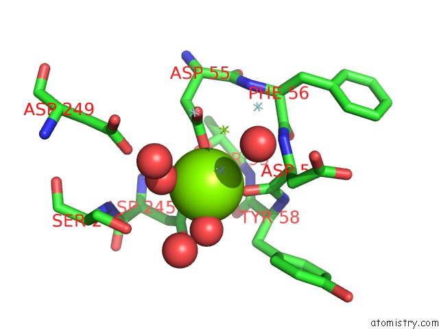
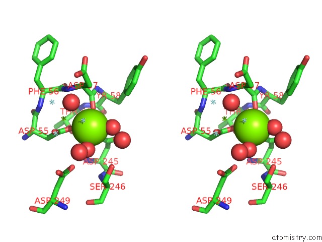
Magnesium binding site 1 out of 4 in 4nv0
Go back to
Magnesium binding site 1 out
of 4 in the Crystal Structure of Cytosolic 5'-Nucleotidase Iiib (Cn-Iiib) Bound to 7-Methylguanosine
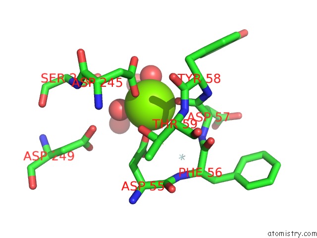
Mono view
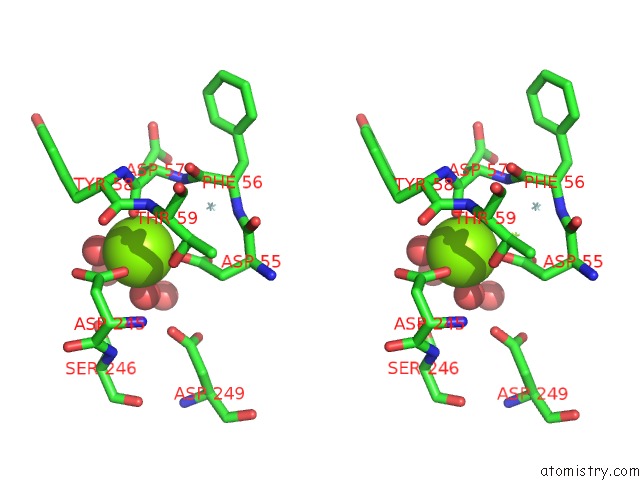
Stereo pair view
Mono view
Stereo pair view
A full contact list of Magnesium with other atoms in the Mg binding
site number 1 of Crystal Structure of Cytosolic 5'-Nucleotidase Iiib (Cn-Iiib) Bound to 7-Methylguanosine within 5.0Å range:
|
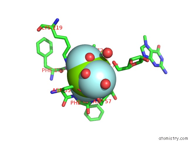
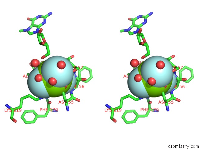
Magnesium binding site 2 out of 4 in 4nv0
Go back to
Magnesium binding site 2 out
of 4 in the Crystal Structure of Cytosolic 5'-Nucleotidase Iiib (Cn-Iiib) Bound to 7-Methylguanosine
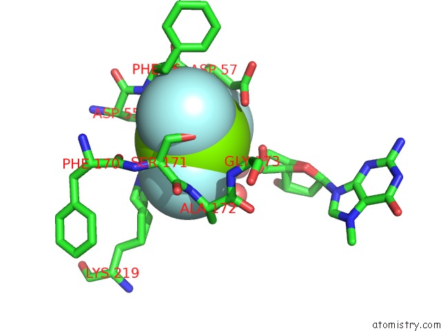
Mono view
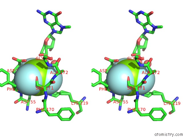
Stereo pair view
Mono view
Stereo pair view
A full contact list of Magnesium with other atoms in the Mg binding
site number 2 of Crystal Structure of Cytosolic 5'-Nucleotidase Iiib (Cn-Iiib) Bound to 7-Methylguanosine within 5.0Å range:
|
Magnesium binding site 3 out of 4 in 4nv0
Go back to
Magnesium binding site 3 out
of 4 in the Crystal Structure of Cytosolic 5'-Nucleotidase Iiib (Cn-Iiib) Bound to 7-Methylguanosine
Mono view
Stereo pair view
Mono view
Stereo pair view
A full contact list of Magnesium with other atoms in the Mg binding
site number 3 of Crystal Structure of Cytosolic 5'-Nucleotidase Iiib (Cn-Iiib) Bound to 7-Methylguanosine within 5.0Å range:
|
Magnesium binding site 4 out of 4 in 4nv0
Go back to
Magnesium binding site 4 out
of 4 in the Crystal Structure of Cytosolic 5'-Nucleotidase Iiib (Cn-Iiib) Bound to 7-Methylguanosine
Mono view
Stereo pair view
Mono view
Stereo pair view
A full contact list of Magnesium with other atoms in the Mg binding
site number 4 of Crystal Structure of Cytosolic 5'-Nucleotidase Iiib (Cn-Iiib) Bound to 7-Methylguanosine within 5.0Å range:
|
Reference:
T.Monecke,
J.Buschmann,
P.Neumann,
E.Wahle,
R.Ficner.
Crystal Structures of the Novel Cytosolic 5'-Nucleotidase Iiib Explain Its Preference For M7GMP Plos One V. 9 90915 2014.
ISSN: ESSN 1932-6203
PubMed: 24603684
DOI: 10.1371/JOURNAL.PONE.0090915
Page generated: Mon Aug 19 23:46:45 2024
ISSN: ESSN 1932-6203
PubMed: 24603684
DOI: 10.1371/JOURNAL.PONE.0090915
Last articles
Zn in 9MJ5Zn in 9HNW
Zn in 9G0L
Zn in 9FNE
Zn in 9DZN
Zn in 9E0I
Zn in 9D32
Zn in 9DAK
Zn in 8ZXC
Zn in 8ZUF