Magnesium »
PDB 4nxn-4o5e »
4nyz »
Magnesium in PDB 4nyz: The Emcv 3DPOL Structure with Altered Motif A Conformation at 2.15A Resolution
Enzymatic activity of The Emcv 3DPOL Structure with Altered Motif A Conformation at 2.15A Resolution
All present enzymatic activity of The Emcv 3DPOL Structure with Altered Motif A Conformation at 2.15A Resolution:
2.7.7.48; 3.4.22.28; 3.6.1.15;
2.7.7.48; 3.4.22.28; 3.6.1.15;
Protein crystallography data
The structure of The Emcv 3DPOL Structure with Altered Motif A Conformation at 2.15A Resolution, PDB code: 4nyz
was solved by
L.Vives-Adrian,
C.Lujan,
C.Ferrer-Orta,
N.Verdaguer,
with X-Ray Crystallography technique. A brief refinement statistics is given in the table below:
| Resolution Low / High (Å) | 104.30 / 2.15 |
| Space group | I 41 2 2 |
| Cell size a, b, c (Å), α, β, γ (°) | 122.553, 122.553, 198.797, 90.00, 90.00, 90.00 |
| R / Rfree (%) | 22.9 / 25.2 |
Magnesium Binding Sites:
The binding sites of Magnesium atom in the The Emcv 3DPOL Structure with Altered Motif A Conformation at 2.15A Resolution
(pdb code 4nyz). This binding sites where shown within
5.0 Angstroms radius around Magnesium atom.
In total only one binding site of Magnesium was determined in the The Emcv 3DPOL Structure with Altered Motif A Conformation at 2.15A Resolution, PDB code: 4nyz:
In total only one binding site of Magnesium was determined in the The Emcv 3DPOL Structure with Altered Motif A Conformation at 2.15A Resolution, PDB code: 4nyz:
Magnesium binding site 1 out of 1 in 4nyz
Go back to
Magnesium binding site 1 out
of 1 in the The Emcv 3DPOL Structure with Altered Motif A Conformation at 2.15A Resolution
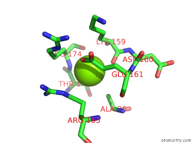
Mono view
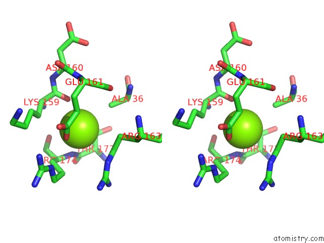
Stereo pair view
Mono view
Stereo pair view
A full contact list of Magnesium with other atoms in the Mg binding
site number 1 of The Emcv 3DPOL Structure with Altered Motif A Conformation at 2.15A Resolution within 5.0Å range:
|
Reference:
L.Vives-Adrian,
C.Lujan,
B.Oliva,
L.Van Der Linden,
B.Selisko,
B.Coutard,
B.Canard,
F.J.Van Kuppeveld,
C.Ferrer-Orta,
N.Verdaguer.
The Crystal Structure of A Cardiovirus Rna-Dependent Rna Polymerase Reveals An Unusual Conformation of the Polymerase Active Site J.Virol. 2014.
ISSN: ESSN 1098-5514
PubMed: 24600002
DOI: 10.1128/JVI.03502-13
Page generated: Tue Aug 20 00:12:59 2024
ISSN: ESSN 1098-5514
PubMed: 24600002
DOI: 10.1128/JVI.03502-13
Last articles
Zn in 9J0NZn in 9J0O
Zn in 9J0P
Zn in 9FJX
Zn in 9EKB
Zn in 9C0F
Zn in 9CAH
Zn in 9CH0
Zn in 9CH3
Zn in 9CH1