Magnesium »
PDB 4nxn-4o5e »
4o2r »
Magnesium in PDB 4o2r: Structure of Mus Musculus Rheb G63V Mutant Bound to Gdp
Protein crystallography data
The structure of Structure of Mus Musculus Rheb G63V Mutant Bound to Gdp, PDB code: 4o2r
was solved by
M.T.Mazhab-Jafari,
C.B.Marshall,
J.Ho,
N.Ishiyama,
V.Stambolic,
M.Ikura,
with X-Ray Crystallography technique. A brief refinement statistics is given in the table below:
| Resolution Low / High (Å) | 28.70 / 2.25 |
| Space group | P 21 21 2 |
| Cell size a, b, c (Å), α, β, γ (°) | 70.334, 79.167, 57.301, 90.00, 90.00, 90.00 |
| R / Rfree (%) | 20.6 / 25.9 |
Magnesium Binding Sites:
The binding sites of Magnesium atom in the Structure of Mus Musculus Rheb G63V Mutant Bound to Gdp
(pdb code 4o2r). This binding sites where shown within
5.0 Angstroms radius around Magnesium atom.
In total 2 binding sites of Magnesium where determined in the Structure of Mus Musculus Rheb G63V Mutant Bound to Gdp, PDB code: 4o2r:
Jump to Magnesium binding site number: 1; 2;
In total 2 binding sites of Magnesium where determined in the Structure of Mus Musculus Rheb G63V Mutant Bound to Gdp, PDB code: 4o2r:
Jump to Magnesium binding site number: 1; 2;
Magnesium binding site 1 out of 2 in 4o2r
Go back to
Magnesium binding site 1 out
of 2 in the Structure of Mus Musculus Rheb G63V Mutant Bound to Gdp
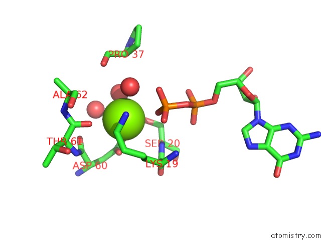
Mono view
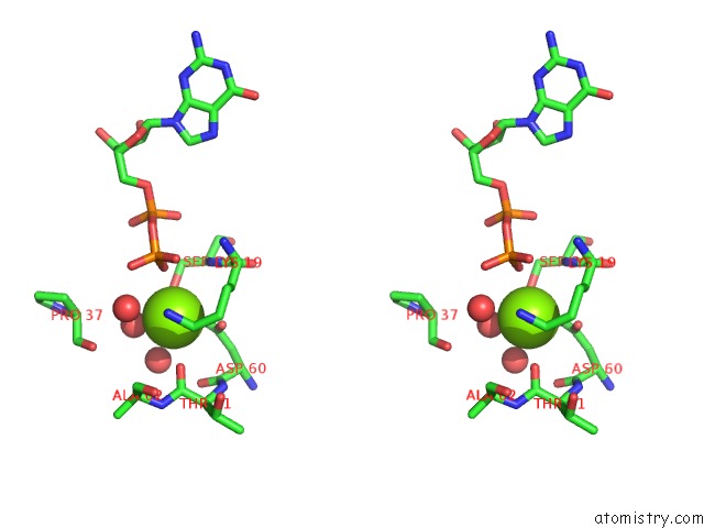
Stereo pair view
Mono view
Stereo pair view
A full contact list of Magnesium with other atoms in the Mg binding
site number 1 of Structure of Mus Musculus Rheb G63V Mutant Bound to Gdp within 5.0Å range:
|
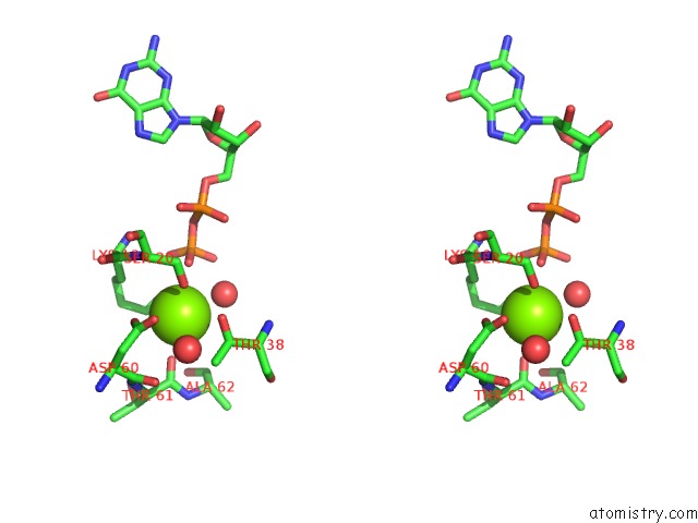
Magnesium binding site 2 out of 2 in 4o2r
Go back to
Magnesium binding site 2 out
of 2 in the Structure of Mus Musculus Rheb G63V Mutant Bound to Gdp
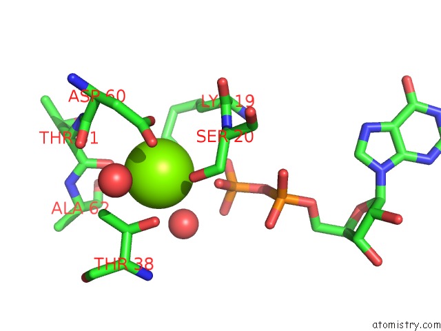
Mono view
Stereo pair view
Mono view
Stereo pair view
A full contact list of Magnesium with other atoms in the Mg binding
site number 2 of Structure of Mus Musculus Rheb G63V Mutant Bound to Gdp within 5.0Å range:
|
Reference:
M.T.Mazhab-Jafari,
C.B.Marshall,
J.Ho,
N.Ishiyama,
V.Stambolic,
M.Ikura.
Structure-Guided Mutation of the Conserved G3-Box Glycine in Rheb Generates A Constitutively Activated Regulator of Mtor To Be Published.
Page generated: Tue Aug 20 00:14:55 2024
Last articles
Zn in 9JYWZn in 9IR4
Zn in 9IR3
Zn in 9GMX
Zn in 9GMW
Zn in 9JEJ
Zn in 9ERF
Zn in 9ERE
Zn in 9EGV
Zn in 9EGW