Magnesium »
PDB 4o5w-4ogu »
4obb »
Magnesium in PDB 4obb: The Crystal Structure of A Solute-Binding Protein From Anabaena Variabilis Atcc 29413 in Complex with (3S)-3-Methyl-2-Oxopentanoic Acid.
Protein crystallography data
The structure of The Crystal Structure of A Solute-Binding Protein From Anabaena Variabilis Atcc 29413 in Complex with (3S)-3-Methyl-2-Oxopentanoic Acid., PDB code: 4obb
was solved by
K.Tan,
H.Li,
R.Jedrzejczak,
A.Joachimiak,
Midwest Center For Structuralgenomics (Mcsg),
with X-Ray Crystallography technique. A brief refinement statistics is given in the table below:
| Resolution Low / High (Å) | 25.86 / 1.53 |
| Space group | C 2 2 21 |
| Cell size a, b, c (Å), α, β, γ (°) | 98.736, 100.814, 150.638, 90.00, 90.00, 90.00 |
| R / Rfree (%) | 13.2 / 17.8 |
Magnesium Binding Sites:
The binding sites of Magnesium atom in the The Crystal Structure of A Solute-Binding Protein From Anabaena Variabilis Atcc 29413 in Complex with (3S)-3-Methyl-2-Oxopentanoic Acid.
(pdb code 4obb). This binding sites where shown within
5.0 Angstroms radius around Magnesium atom.
In total 2 binding sites of Magnesium where determined in the The Crystal Structure of A Solute-Binding Protein From Anabaena Variabilis Atcc 29413 in Complex with (3S)-3-Methyl-2-Oxopentanoic Acid., PDB code: 4obb:
Jump to Magnesium binding site number: 1; 2;
In total 2 binding sites of Magnesium where determined in the The Crystal Structure of A Solute-Binding Protein From Anabaena Variabilis Atcc 29413 in Complex with (3S)-3-Methyl-2-Oxopentanoic Acid., PDB code: 4obb:
Jump to Magnesium binding site number: 1; 2;
Magnesium binding site 1 out of 2 in 4obb
Go back to
Magnesium binding site 1 out
of 2 in the The Crystal Structure of A Solute-Binding Protein From Anabaena Variabilis Atcc 29413 in Complex with (3S)-3-Methyl-2-Oxopentanoic Acid.
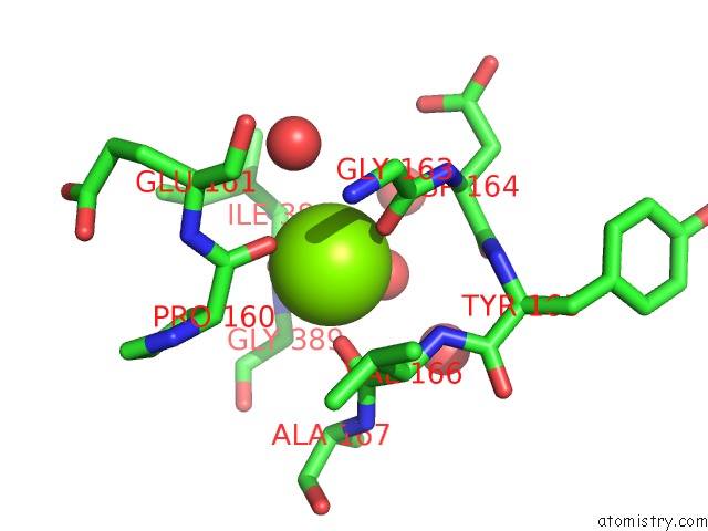
Mono view
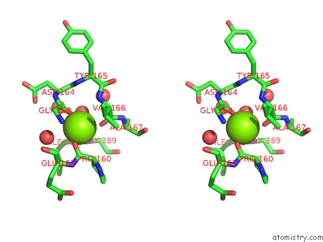
Stereo pair view
Mono view
Stereo pair view
A full contact list of Magnesium with other atoms in the Mg binding
site number 1 of The Crystal Structure of A Solute-Binding Protein From Anabaena Variabilis Atcc 29413 in Complex with (3S)-3-Methyl-2-Oxopentanoic Acid. within 5.0Å range:
|
Magnesium binding site 2 out of 2 in 4obb
Go back to
Magnesium binding site 2 out
of 2 in the The Crystal Structure of A Solute-Binding Protein From Anabaena Variabilis Atcc 29413 in Complex with (3S)-3-Methyl-2-Oxopentanoic Acid.
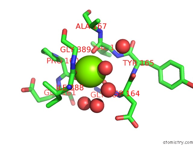
Mono view
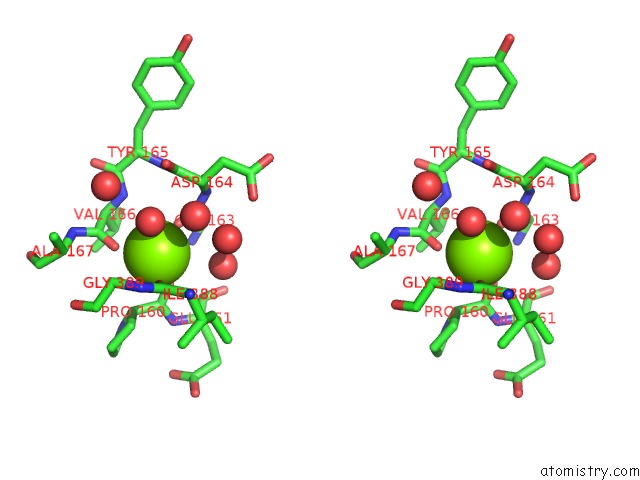
Stereo pair view
Mono view
Stereo pair view
A full contact list of Magnesium with other atoms in the Mg binding
site number 2 of The Crystal Structure of A Solute-Binding Protein From Anabaena Variabilis Atcc 29413 in Complex with (3S)-3-Methyl-2-Oxopentanoic Acid. within 5.0Å range:
|
Reference:
K.Tan,
H.Li,
R.Jedrzejczak,
A.Joachimiak.
The Crystal Structure of A Solute-Binding Protein From Anabaena Variabilis Atcc 29413 in Complex with (3S)-3-Methyl-2-Oxopentanoic Acid. To Be Published.
Page generated: Mon Aug 11 21:27:17 2025
Last articles
Mg in 6HNQMg in 6HOS
Mg in 6HNS
Mg in 6HN2
Mg in 6HMZ
Mg in 6HMU
Mg in 6HMT
Mg in 6HLR
Mg in 6HLQ
Mg in 6HKY