Magnesium »
PDB 4o5w-4ogu »
4odj »
Magnesium in PDB 4odj: Crystal Structure of A Putative S-Adenosylmethionine Synthetase From Cryptosporidium Hominis in Complex with S-Adenosyl-Methionine
Enzymatic activity of Crystal Structure of A Putative S-Adenosylmethionine Synthetase From Cryptosporidium Hominis in Complex with S-Adenosyl-Methionine
All present enzymatic activity of Crystal Structure of A Putative S-Adenosylmethionine Synthetase From Cryptosporidium Hominis in Complex with S-Adenosyl-Methionine:
2.5.1.6;
2.5.1.6;
Protein crystallography data
The structure of Crystal Structure of A Putative S-Adenosylmethionine Synthetase From Cryptosporidium Hominis in Complex with S-Adenosyl-Methionine, PDB code: 4odj
was solved by
Seattle Structural Gemomics Center For Infectious Disease (Ssgcid),
with X-Ray Crystallography technique. A brief refinement statistics is given in the table below:
| Resolution Low / High (Å) | 50.00 / 1.60 |
| Space group | P 32 2 1 |
| Cell size a, b, c (Å), α, β, γ (°) | 117.860, 117.860, 54.040, 90.00, 90.00, 120.00 |
| R / Rfree (%) | 14.4 / 16.5 |
Magnesium Binding Sites:
The binding sites of Magnesium atom in the Crystal Structure of A Putative S-Adenosylmethionine Synthetase From Cryptosporidium Hominis in Complex with S-Adenosyl-Methionine
(pdb code 4odj). This binding sites where shown within
5.0 Angstroms radius around Magnesium atom.
In total 2 binding sites of Magnesium where determined in the Crystal Structure of A Putative S-Adenosylmethionine Synthetase From Cryptosporidium Hominis in Complex with S-Adenosyl-Methionine, PDB code: 4odj:
Jump to Magnesium binding site number: 1; 2;
In total 2 binding sites of Magnesium where determined in the Crystal Structure of A Putative S-Adenosylmethionine Synthetase From Cryptosporidium Hominis in Complex with S-Adenosyl-Methionine, PDB code: 4odj:
Jump to Magnesium binding site number: 1; 2;
Magnesium binding site 1 out of 2 in 4odj
Go back to
Magnesium binding site 1 out
of 2 in the Crystal Structure of A Putative S-Adenosylmethionine Synthetase From Cryptosporidium Hominis in Complex with S-Adenosyl-Methionine
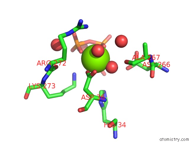
Mono view
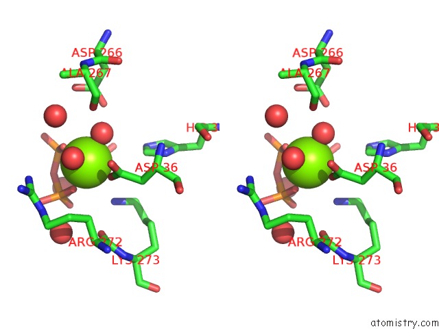
Stereo pair view
Mono view
Stereo pair view
A full contact list of Magnesium with other atoms in the Mg binding
site number 1 of Crystal Structure of A Putative S-Adenosylmethionine Synthetase From Cryptosporidium Hominis in Complex with S-Adenosyl-Methionine within 5.0Å range:
|
Magnesium binding site 2 out of 2 in 4odj
Go back to
Magnesium binding site 2 out
of 2 in the Crystal Structure of A Putative S-Adenosylmethionine Synthetase From Cryptosporidium Hominis in Complex with S-Adenosyl-Methionine
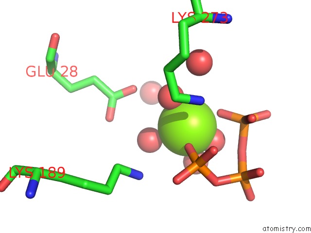
Mono view
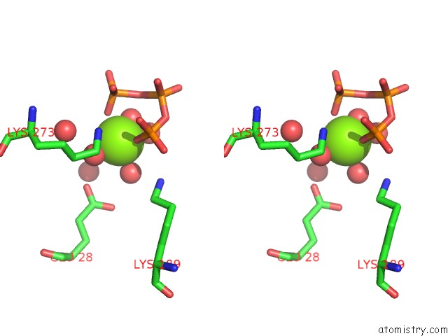
Stereo pair view
Mono view
Stereo pair view
A full contact list of Magnesium with other atoms in the Mg binding
site number 2 of Crystal Structure of A Putative S-Adenosylmethionine Synthetase From Cryptosporidium Hominis in Complex with S-Adenosyl-Methionine within 5.0Å range:
|
Reference:
Seattle Structural Genomics Center For Infectious Disease(Ssgcid),
J.Abendroth,
T.Arakaki,
D.Lorimer,
T.E.Edewars.
Crystal Structure of A Putative S-Adenosylmethionine Synthetase From Cryptosporidium Hominis in Complex with S-Adenosyl-Methionine To Be Published.
Page generated: Mon Aug 11 21:28:09 2025
Last articles
Mg in 5H9BMg in 5H8Y
Mg in 5H92
Mg in 5H8V
Mg in 5H8U
Mg in 5H7O
Mg in 5H5X
Mg in 5H74
Mg in 5H8M
Mg in 5H56