Magnesium »
PDB 4o5w-4ogu »
4ofe »
Magnesium in PDB 4ofe: Structural Basis For Thymine Glycosylase Activity on T:O6-Methylg Mismatch By Methyl-Cpg Binding Domain Protein 4: Implications For Roles of ARG468 in Mismatch Recognition and Catalysis
Protein crystallography data
The structure of Structural Basis For Thymine Glycosylase Activity on T:O6-Methylg Mismatch By Methyl-Cpg Binding Domain Protein 4: Implications For Roles of ARG468 in Mismatch Recognition and Catalysis, PDB code: 4ofe
was solved by
H.Ouzon-Shubeita,
Y.-L.Lin,
S.Lee,
with X-Ray Crystallography technique. A brief refinement statistics is given in the table below:
| Resolution Low / High (Å) | 33.39 / 2.15 |
| Space group | P 21 21 21 |
| Cell size a, b, c (Å), α, β, γ (°) | 41.722, 55.605, 104.245, 90.00, 90.00, 90.00 |
| R / Rfree (%) | 14.2 / 20.2 |
Magnesium Binding Sites:
The binding sites of Magnesium atom in the Structural Basis For Thymine Glycosylase Activity on T:O6-Methylg Mismatch By Methyl-Cpg Binding Domain Protein 4: Implications For Roles of ARG468 in Mismatch Recognition and Catalysis
(pdb code 4ofe). This binding sites where shown within
5.0 Angstroms radius around Magnesium atom.
In total only one binding site of Magnesium was determined in the Structural Basis For Thymine Glycosylase Activity on T:O6-Methylg Mismatch By Methyl-Cpg Binding Domain Protein 4: Implications For Roles of ARG468 in Mismatch Recognition and Catalysis, PDB code: 4ofe:
In total only one binding site of Magnesium was determined in the Structural Basis For Thymine Glycosylase Activity on T:O6-Methylg Mismatch By Methyl-Cpg Binding Domain Protein 4: Implications For Roles of ARG468 in Mismatch Recognition and Catalysis, PDB code: 4ofe:
Magnesium binding site 1 out of 1 in 4ofe
Go back to
Magnesium binding site 1 out
of 1 in the Structural Basis For Thymine Glycosylase Activity on T:O6-Methylg Mismatch By Methyl-Cpg Binding Domain Protein 4: Implications For Roles of ARG468 in Mismatch Recognition and Catalysis
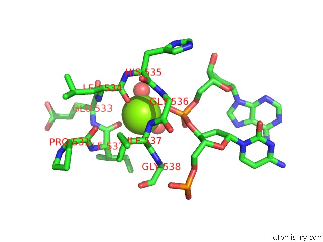
Mono view
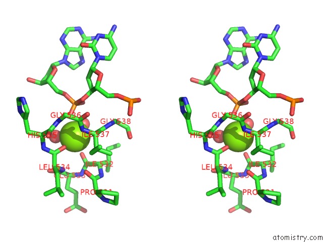
Stereo pair view
Mono view
Stereo pair view
A full contact list of Magnesium with other atoms in the Mg binding
site number 1 of Structural Basis For Thymine Glycosylase Activity on T:O6-Methylg Mismatch By Methyl-Cpg Binding Domain Protein 4: Implications For Roles of ARG468 in Mismatch Recognition and Catalysis within 5.0Å range:
|
Reference:
H.Ouzon-Shubeita,
Y.-L.Lin,
S.Lee.
Structure of R468K/D560N MBD4 Bound to G:T Mispair Dna To Be Published.
Page generated: Mon Aug 11 21:29:48 2025
Last articles
Mg in 5DS5Mg in 5DRZ
Mg in 5DRI
Mg in 5DRC
Mg in 5DRD
Mg in 5DQL
Mg in 5DR2
Mg in 5DQZ
Mg in 5DQK
Mg in 5DOU