Magnesium »
PDB 4o5w-4ogu »
4og2 »
Magnesium in PDB 4og2: The Crystal Structure of A Solute-Binding Protein (N280D Mutant) From Anabaena Variabilis Atcc 29413 in Complex with Leucine
Protein crystallography data
The structure of The Crystal Structure of A Solute-Binding Protein (N280D Mutant) From Anabaena Variabilis Atcc 29413 in Complex with Leucine, PDB code: 4og2
was solved by
K.Tan,
H.Li,
R.Jedrzejczak,
A.Joachimiak,
Midwest Center For Structuralgenomics (Mcsg),
with X-Ray Crystallography technique. A brief refinement statistics is given in the table below:
| Resolution Low / High (Å) | 18.50 / 1.10 |
| Space group | C 2 2 21 |
| Cell size a, b, c (Å), α, β, γ (°) | 98.326, 101.124, 149.967, 90.00, 90.00, 90.00 |
| R / Rfree (%) | 12.9 / 15 |
Other elements in 4og2:
The structure of The Crystal Structure of A Solute-Binding Protein (N280D Mutant) From Anabaena Variabilis Atcc 29413 in Complex with Leucine also contains other interesting chemical elements:
| Chlorine | (Cl) | 2 atoms |
Magnesium Binding Sites:
The binding sites of Magnesium atom in the The Crystal Structure of A Solute-Binding Protein (N280D Mutant) From Anabaena Variabilis Atcc 29413 in Complex with Leucine
(pdb code 4og2). This binding sites where shown within
5.0 Angstroms radius around Magnesium atom.
In total 2 binding sites of Magnesium where determined in the The Crystal Structure of A Solute-Binding Protein (N280D Mutant) From Anabaena Variabilis Atcc 29413 in Complex with Leucine, PDB code: 4og2:
Jump to Magnesium binding site number: 1; 2;
In total 2 binding sites of Magnesium where determined in the The Crystal Structure of A Solute-Binding Protein (N280D Mutant) From Anabaena Variabilis Atcc 29413 in Complex with Leucine, PDB code: 4og2:
Jump to Magnesium binding site number: 1; 2;
Magnesium binding site 1 out of 2 in 4og2
Go back to
Magnesium binding site 1 out
of 2 in the The Crystal Structure of A Solute-Binding Protein (N280D Mutant) From Anabaena Variabilis Atcc 29413 in Complex with Leucine
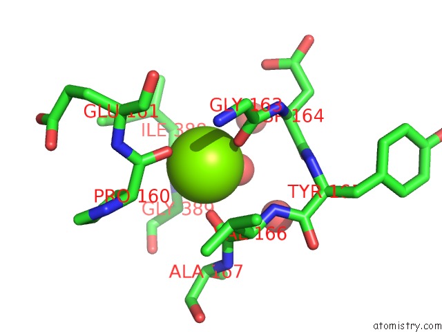
Mono view
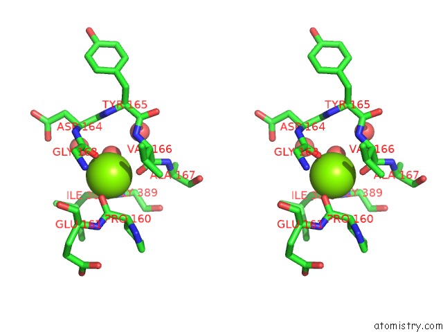
Stereo pair view
Mono view
Stereo pair view
A full contact list of Magnesium with other atoms in the Mg binding
site number 1 of The Crystal Structure of A Solute-Binding Protein (N280D Mutant) From Anabaena Variabilis Atcc 29413 in Complex with Leucine within 5.0Å range:
|
Magnesium binding site 2 out of 2 in 4og2
Go back to
Magnesium binding site 2 out
of 2 in the The Crystal Structure of A Solute-Binding Protein (N280D Mutant) From Anabaena Variabilis Atcc 29413 in Complex with Leucine

Mono view

Stereo pair view
Mono view
Stereo pair view
A full contact list of Magnesium with other atoms in the Mg binding
site number 2 of The Crystal Structure of A Solute-Binding Protein (N280D Mutant) From Anabaena Variabilis Atcc 29413 in Complex with Leucine within 5.0Å range:
|
Reference:
K.Tan,
H.Li,
R.Jedrzejczak,
A.Joachimiak.
The Crystal Structure of A Solute-Binding Protein (N280D Mutant) From Anabaena Variabilis Atcc 29413 in Complex with Leucine To Be Published.
Page generated: Tue Aug 20 00:49:36 2024
Last articles
F in 4HGSF in 4H52
F in 4H4O
F in 4H4C
F in 4GSY
F in 4H3J
F in 4GXJ
F in 4GVH
F in 4GU9
F in 4GSI