Magnesium »
PDB 4ohf-4osl »
4okj »
Magnesium in PDB 4okj: Crystal Structure of Rnase As From M Tuberculosis
Protein crystallography data
The structure of Crystal Structure of Rnase As From M Tuberculosis, PDB code: 4okj
was solved by
M.Romano,
R.Van De Weerd,
F.C.C.Brouwer,
G.N.Roviello,
R.Lacroix,
M.Sparrius,
G.Van Den Brink-Van Stempvoort,
J.J.Maaskant,
A.M.Van Dersar,
B.J.Appelmelk,
J.J.Geurtsen,
R.Berisio,
with X-Ray Crystallography technique. A brief refinement statistics is given in the table below:
| Resolution Low / High (Å) | 14.89 / 2.10 |
| Space group | P 21 21 21 |
| Cell size a, b, c (Å), α, β, γ (°) | 42.424, 77.055, 104.580, 90.00, 90.00, 90.00 |
| R / Rfree (%) | 18.5 / 25.7 |
Magnesium Binding Sites:
The binding sites of Magnesium atom in the Crystal Structure of Rnase As From M Tuberculosis
(pdb code 4okj). This binding sites where shown within
5.0 Angstroms radius around Magnesium atom.
In total 6 binding sites of Magnesium where determined in the Crystal Structure of Rnase As From M Tuberculosis, PDB code: 4okj:
Jump to Magnesium binding site number: 1; 2; 3; 4; 5; 6;
In total 6 binding sites of Magnesium where determined in the Crystal Structure of Rnase As From M Tuberculosis, PDB code: 4okj:
Jump to Magnesium binding site number: 1; 2; 3; 4; 5; 6;
Magnesium binding site 1 out of 6 in 4okj
Go back to
Magnesium binding site 1 out
of 6 in the Crystal Structure of Rnase As From M Tuberculosis

Mono view
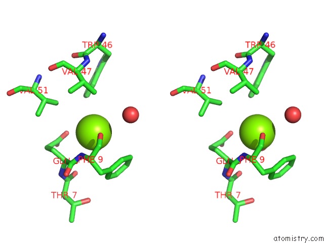
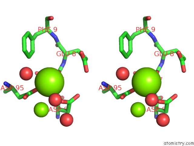
Stereo pair view
Mono view
Stereo pair view
A full contact list of Magnesium with other atoms in the Mg binding
site number 1 of Crystal Structure of Rnase As From M Tuberculosis within 5.0Å range:
|
Magnesium binding site 2 out of 6 in 4okj
Go back to
Magnesium binding site 2 out
of 6 in the Crystal Structure of Rnase As From M Tuberculosis
Mono view
Stereo pair view
Mono view
Stereo pair view
A full contact list of Magnesium with other atoms in the Mg binding
site number 2 of Crystal Structure of Rnase As From M Tuberculosis within 5.0Å range:
|
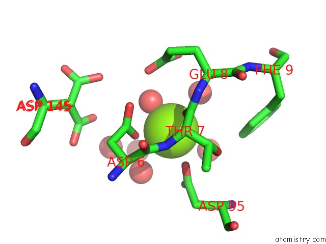
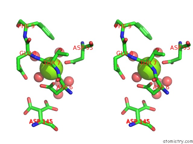
Magnesium binding site 3 out of 6 in 4okj
Go back to
Magnesium binding site 3 out
of 6 in the Crystal Structure of Rnase As From M Tuberculosis

Mono view

Stereo pair view
Mono view
Stereo pair view
A full contact list of Magnesium with other atoms in the Mg binding
site number 3 of Crystal Structure of Rnase As From M Tuberculosis within 5.0Å range:
|
Magnesium binding site 4 out of 6 in 4okj
Go back to
Magnesium binding site 4 out
of 6 in the Crystal Structure of Rnase As From M Tuberculosis
Mono view

Stereo pair view
Mono view
Stereo pair view
A full contact list of Magnesium with other atoms in the Mg binding
site number 4 of Crystal Structure of Rnase As From M Tuberculosis within 5.0Å range:
|
Magnesium binding site 5 out of 6 in 4okj
Go back to
Magnesium binding site 5 out
of 6 in the Crystal Structure of Rnase As From M Tuberculosis

Mono view
Stereo pair view
Mono view
Stereo pair view
A full contact list of Magnesium with other atoms in the Mg binding
site number 5 of Crystal Structure of Rnase As From M Tuberculosis within 5.0Å range:
|
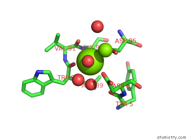
Magnesium binding site 6 out of 6 in 4okj
Go back to
Magnesium binding site 6 out
of 6 in the Crystal Structure of Rnase As From M Tuberculosis

Mono view

Stereo pair view
Mono view
Stereo pair view
A full contact list of Magnesium with other atoms in the Mg binding
site number 6 of Crystal Structure of Rnase As From M Tuberculosis within 5.0Å range:
|
Reference:
M.Romano,
R.Van De Weerd,
F.C.C.Brouwer,
G.N.Roviello,
R.Lacroix,
M.Sparrius,
G.Van Den Brink-Van Stempvoort,
J.J.Maaskant,
A.M.Van Der Sar,
B.J.Appelmelk,
J.J.Geurtsen,
R.Berisio.
Structure and Function of Rnase As, A Polyadenylate-Specific Exoribonuclease Affecting Mycobacterial Virulence in Vivo Structure V. 22 719 2014.
ISSN: ISSN 0969-2126
PubMed: 24704253
DOI: 10.1016/J.STR.2014.01.014
Page generated: Tue Aug 20 00:53:39 2024
ISSN: ISSN 0969-2126
PubMed: 24704253
DOI: 10.1016/J.STR.2014.01.014
Last articles
Zn in 9MJ5Zn in 9HNW
Zn in 9G0L
Zn in 9FNE
Zn in 9DZN
Zn in 9E0I
Zn in 9D32
Zn in 9DAK
Zn in 8ZXC
Zn in 8ZUF