Magnesium »
PDB 4osl-4p7a »
4ott »
Magnesium in PDB 4ott: Crystal Structure of the Gamma-Glutamyltranspeptidase From Bacillus Licheniformis.
Enzymatic activity of Crystal Structure of the Gamma-Glutamyltranspeptidase From Bacillus Licheniformis.
All present enzymatic activity of Crystal Structure of the Gamma-Glutamyltranspeptidase From Bacillus Licheniformis.:
2.3.2.2;
2.3.2.2;
Protein crystallography data
The structure of Crystal Structure of the Gamma-Glutamyltranspeptidase From Bacillus Licheniformis., PDB code: 4ott
was solved by
A.Merlino,
with X-Ray Crystallography technique. A brief refinement statistics is given in the table below:
| Resolution Low / High (Å) | 26.25 / 2.98 |
| Space group | P 21 21 21 |
| Cell size a, b, c (Å), α, β, γ (°) | 60.896, 61.968, 148.245, 90.00, 90.00, 90.00 |
| R / Rfree (%) | 19.1 / 25 |
Magnesium Binding Sites:
The binding sites of Magnesium atom in the Crystal Structure of the Gamma-Glutamyltranspeptidase From Bacillus Licheniformis.
(pdb code 4ott). This binding sites where shown within
5.0 Angstroms radius around Magnesium atom.
In total 2 binding sites of Magnesium where determined in the Crystal Structure of the Gamma-Glutamyltranspeptidase From Bacillus Licheniformis., PDB code: 4ott:
Jump to Magnesium binding site number: 1; 2;
In total 2 binding sites of Magnesium where determined in the Crystal Structure of the Gamma-Glutamyltranspeptidase From Bacillus Licheniformis., PDB code: 4ott:
Jump to Magnesium binding site number: 1; 2;
Magnesium binding site 1 out of 2 in 4ott
Go back to
Magnesium binding site 1 out
of 2 in the Crystal Structure of the Gamma-Glutamyltranspeptidase From Bacillus Licheniformis.
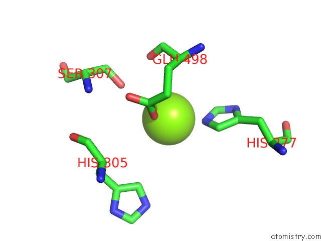
Mono view
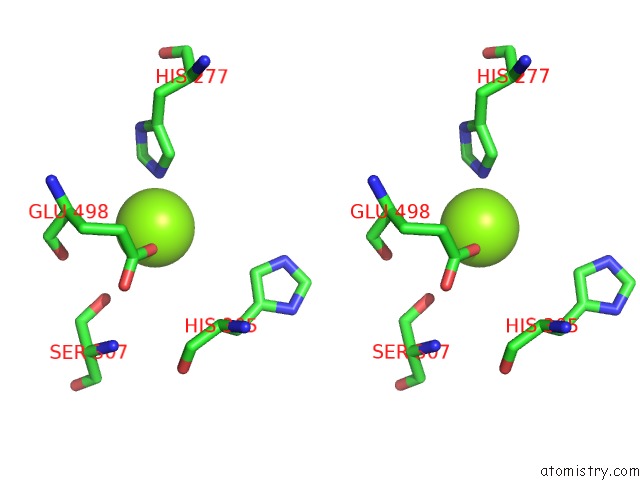
Stereo pair view
Mono view
Stereo pair view
A full contact list of Magnesium with other atoms in the Mg binding
site number 1 of Crystal Structure of the Gamma-Glutamyltranspeptidase From Bacillus Licheniformis. within 5.0Å range:
|
Magnesium binding site 2 out of 2 in 4ott
Go back to
Magnesium binding site 2 out
of 2 in the Crystal Structure of the Gamma-Glutamyltranspeptidase From Bacillus Licheniformis.
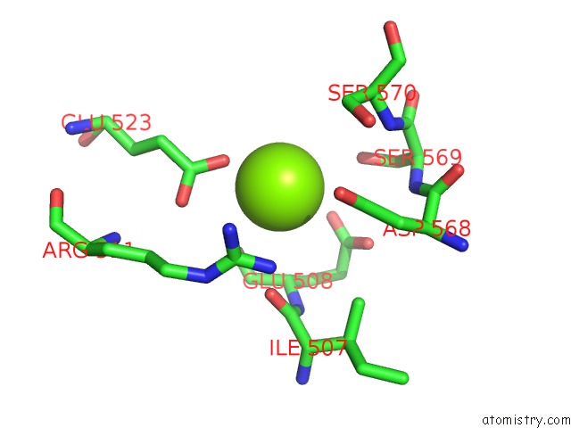
Mono view
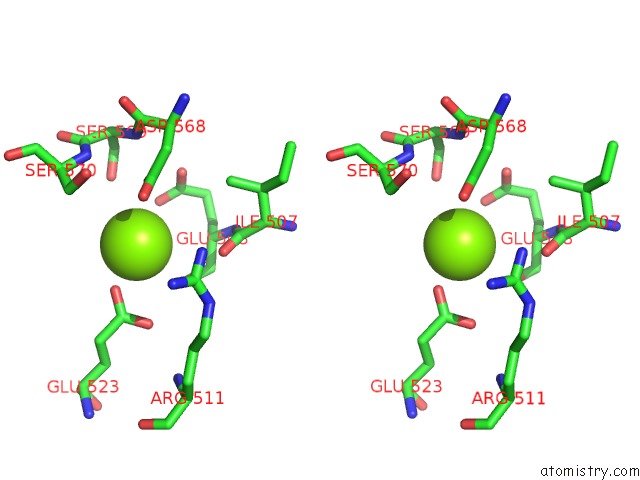
Stereo pair view
Mono view
Stereo pair view
A full contact list of Magnesium with other atoms in the Mg binding
site number 2 of Crystal Structure of the Gamma-Glutamyltranspeptidase From Bacillus Licheniformis. within 5.0Å range:
|
Reference:
L.L.Lin,
Y.Y.Chen,
M.C.Chi,
A.Merlino.
Low Resolution X-Ray Structure of Gamma-Glutamyltranspeptidase From Bacillus Licheniformis: Opened Active Site Cleft and A Cluster of Acid Residues Potentially Involved in the Recognition of A Metal Ion. Biochim.Biophys.Acta V.1844 1523 2014.
ISSN: ISSN 0006-3002
PubMed: 24780583
DOI: 10.1016/J.BBAPAP.2014.04.016
Page generated: Tue Aug 20 00:59:39 2024
ISSN: ISSN 0006-3002
PubMed: 24780583
DOI: 10.1016/J.BBAPAP.2014.04.016
Last articles
Zn in 9J0NZn in 9J0O
Zn in 9J0P
Zn in 9FJX
Zn in 9EKB
Zn in 9C0F
Zn in 9CAH
Zn in 9CH0
Zn in 9CH3
Zn in 9CH1