Magnesium »
PDB 4osq-4p8r »
4p8r »
Magnesium in PDB 4p8r: Structure of A Glycosomal Glyceraldehyde 3-Phosphate Dehydrogenase From Trypanosoma Brucei
Enzymatic activity of Structure of A Glycosomal Glyceraldehyde 3-Phosphate Dehydrogenase From Trypanosoma Brucei
All present enzymatic activity of Structure of A Glycosomal Glyceraldehyde 3-Phosphate Dehydrogenase From Trypanosoma Brucei:
1.2.1.12;
1.2.1.12;
Protein crystallography data
The structure of Structure of A Glycosomal Glyceraldehyde 3-Phosphate Dehydrogenase From Trypanosoma Brucei, PDB code: 4p8r
was solved by
Seattle Structural Genomics Center For Infectious Disease (Ssgcid),
with X-Ray Crystallography technique. A brief refinement statistics is given in the table below:
| Resolution Low / High (Å) | 19.89 / 2.20 |
| Space group | P 65 2 2 |
| Cell size a, b, c (Å), α, β, γ (°) | 86.700, 86.700, 701.370, 90.00, 90.00, 120.00 |
| R / Rfree (%) | 15 / 19.7 |
Magnesium Binding Sites:
The binding sites of Magnesium atom in the Structure of A Glycosomal Glyceraldehyde 3-Phosphate Dehydrogenase From Trypanosoma Brucei
(pdb code 4p8r). This binding sites where shown within
5.0 Angstroms radius around Magnesium atom.
In total 3 binding sites of Magnesium where determined in the Structure of A Glycosomal Glyceraldehyde 3-Phosphate Dehydrogenase From Trypanosoma Brucei, PDB code: 4p8r:
Jump to Magnesium binding site number: 1; 2; 3;
In total 3 binding sites of Magnesium where determined in the Structure of A Glycosomal Glyceraldehyde 3-Phosphate Dehydrogenase From Trypanosoma Brucei, PDB code: 4p8r:
Jump to Magnesium binding site number: 1; 2; 3;
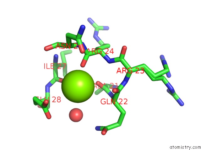
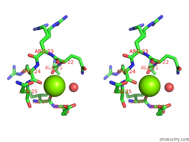
Magnesium binding site 1 out of 3 in 4p8r
Go back to
Magnesium binding site 1 out
of 3 in the Structure of A Glycosomal Glyceraldehyde 3-Phosphate Dehydrogenase From Trypanosoma Brucei
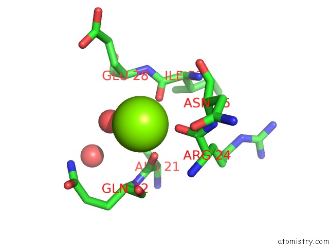
Mono view
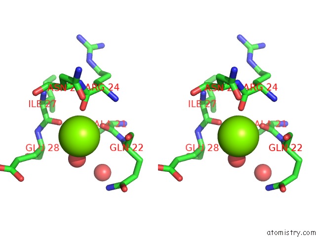
Stereo pair view
Mono view
Stereo pair view
A full contact list of Magnesium with other atoms in the Mg binding
site number 1 of Structure of A Glycosomal Glyceraldehyde 3-Phosphate Dehydrogenase From Trypanosoma Brucei within 5.0Å range:
|
Magnesium binding site 2 out of 3 in 4p8r
Go back to
Magnesium binding site 2 out
of 3 in the Structure of A Glycosomal Glyceraldehyde 3-Phosphate Dehydrogenase From Trypanosoma Brucei
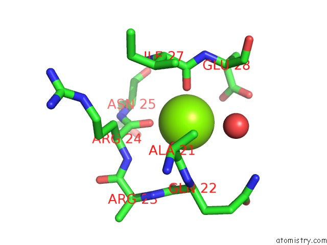
Mono view
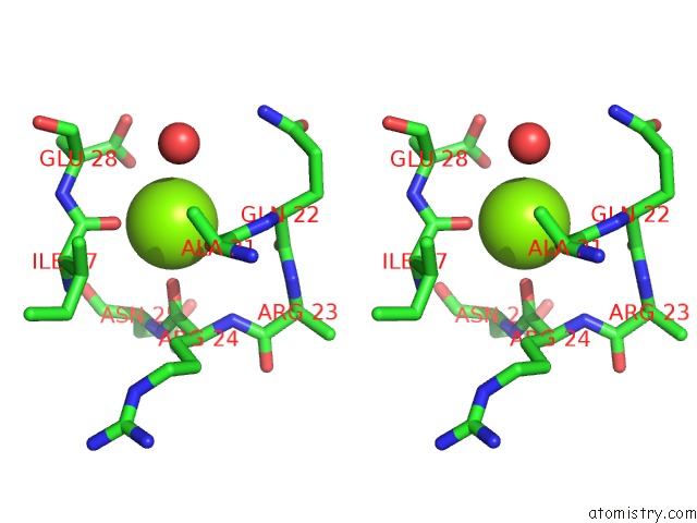
Stereo pair view
Mono view
Stereo pair view
A full contact list of Magnesium with other atoms in the Mg binding
site number 2 of Structure of A Glycosomal Glyceraldehyde 3-Phosphate Dehydrogenase From Trypanosoma Brucei within 5.0Å range:
|
Magnesium binding site 3 out of 3 in 4p8r
Go back to
Magnesium binding site 3 out
of 3 in the Structure of A Glycosomal Glyceraldehyde 3-Phosphate Dehydrogenase From Trypanosoma Brucei
Mono view
Stereo pair view
Mono view
Stereo pair view
A full contact list of Magnesium with other atoms in the Mg binding
site number 3 of Structure of A Glycosomal Glyceraldehyde 3-Phosphate Dehydrogenase From Trypanosoma Brucei within 5.0Å range:
|
Reference:
Seattle Structural Genomics Center For Infectious Disease(Ssgcid),
J.Abendroth,
D.Lorimer,
T.E.Edwards.
Structure of A Glycosomal Glyceraldehyde 3-Phosphate Dehydrogenase From Trypanosoma Brucei To Be Published.
Page generated: Mon Aug 11 21:44:04 2025
Last articles
Mg in 5SBEMg in 5SBD
Mg in 5SBC
Mg in 5SBB
Mg in 5SBA
Mg in 5SB8
Mg in 5SB9
Mg in 5SB7
Mg in 5SB6
Mg in 5SB4