Magnesium »
PDB 4p9d-4pjj »
4pht »
Magnesium in PDB 4pht: Atpase Gspe in Complex with the Cytoplasmic Domain of Gspl From the Vibrio Vulnificus Type II Secretion System
Protein crystallography data
The structure of Atpase Gspe in Complex with the Cytoplasmic Domain of Gspl From the Vibrio Vulnificus Type II Secretion System, PDB code: 4pht
was solved by
C.Lu,
K.Korotkov,
W.Hol,
with X-Ray Crystallography technique. A brief refinement statistics is given in the table below:
| Resolution Low / High (Å) | 43.22 / 2.83 |
| Space group | C 1 2 1 |
| Cell size a, b, c (Å), α, β, γ (°) | 226.430, 133.900, 93.490, 90.00, 91.41, 90.00 |
| R / Rfree (%) | 24.8 / 28 |
Other elements in 4pht:
The structure of Atpase Gspe in Complex with the Cytoplasmic Domain of Gspl From the Vibrio Vulnificus Type II Secretion System also contains other interesting chemical elements:
| Zinc | (Zn) | 3 atoms |
Magnesium Binding Sites:
The binding sites of Magnesium atom in the Atpase Gspe in Complex with the Cytoplasmic Domain of Gspl From the Vibrio Vulnificus Type II Secretion System
(pdb code 4pht). This binding sites where shown within
5.0 Angstroms radius around Magnesium atom.
In total 3 binding sites of Magnesium where determined in the Atpase Gspe in Complex with the Cytoplasmic Domain of Gspl From the Vibrio Vulnificus Type II Secretion System, PDB code: 4pht:
Jump to Magnesium binding site number: 1; 2; 3;
In total 3 binding sites of Magnesium where determined in the Atpase Gspe in Complex with the Cytoplasmic Domain of Gspl From the Vibrio Vulnificus Type II Secretion System, PDB code: 4pht:
Jump to Magnesium binding site number: 1; 2; 3;
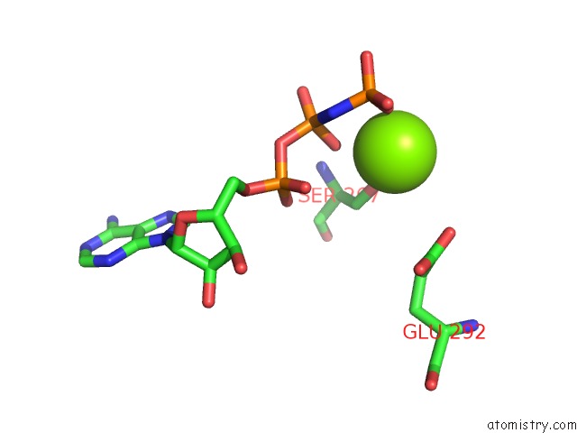
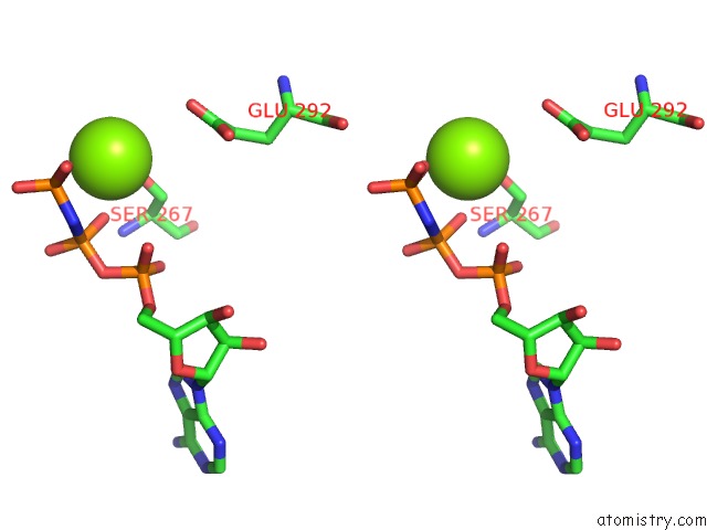
Magnesium binding site 1 out of 3 in 4pht
Go back to
Magnesium binding site 1 out
of 3 in the Atpase Gspe in Complex with the Cytoplasmic Domain of Gspl From the Vibrio Vulnificus Type II Secretion System

Mono view

Stereo pair view
Mono view
Stereo pair view
A full contact list of Magnesium with other atoms in the Mg binding
site number 1 of Atpase Gspe in Complex with the Cytoplasmic Domain of Gspl From the Vibrio Vulnificus Type II Secretion System within 5.0Å range:
|
Magnesium binding site 2 out of 3 in 4pht
Go back to
Magnesium binding site 2 out
of 3 in the Atpase Gspe in Complex with the Cytoplasmic Domain of Gspl From the Vibrio Vulnificus Type II Secretion System
Mono view
Stereo pair view
Mono view
Stereo pair view
A full contact list of Magnesium with other atoms in the Mg binding
site number 2 of Atpase Gspe in Complex with the Cytoplasmic Domain of Gspl From the Vibrio Vulnificus Type II Secretion System within 5.0Å range:
|
Magnesium binding site 3 out of 3 in 4pht
Go back to
Magnesium binding site 3 out
of 3 in the Atpase Gspe in Complex with the Cytoplasmic Domain of Gspl From the Vibrio Vulnificus Type II Secretion System

Mono view

Stereo pair view
Mono view
Stereo pair view
A full contact list of Magnesium with other atoms in the Mg binding
site number 3 of Atpase Gspe in Complex with the Cytoplasmic Domain of Gspl From the Vibrio Vulnificus Type II Secretion System within 5.0Å range:
|
Reference:
C.Lu,
K.V.Korotkov,
W.G.Hol.
Crystal Structure of the Full-Length Atpase Gspe From the Vibrio Vulnificus Type II Secretion System in Complex with the Cytoplasmic Domain of Gspl. J.Struct.Biol. V. 187 223 2014.
ISSN: ESSN 1095-8657
PubMed: 25092625
DOI: 10.1016/J.JSB.2014.07.006
Page generated: Mon Aug 11 21:55:21 2025
ISSN: ESSN 1095-8657
PubMed: 25092625
DOI: 10.1016/J.JSB.2014.07.006
Last articles
Mg in 5EX2Mg in 5EX1
Mg in 5EW7
Mg in 5EW4
Mg in 5EVZ
Mg in 5ETS
Mg in 5ETT
Mg in 5EUR
Mg in 5EUL
Mg in 5ETV