Magnesium »
PDB 4pyk-4q86 »
4q4y »
Magnesium in PDB 4q4y: Crystal Structure of Coxsackievirus A24V Soaked with Disialyllacto-N- Tetraose (Dslnt)
Protein crystallography data
The structure of Crystal Structure of Coxsackievirus A24V Soaked with Disialyllacto-N- Tetraose (Dslnt), PDB code: 4q4y
was solved by
G.Zocher,
T.Stehle,
with X-Ray Crystallography technique. A brief refinement statistics is given in the table below:
| Resolution Low / High (Å) | 49.78 / 1.88 |
| Space group | I 2 2 2 |
| Cell size a, b, c (Å), α, β, γ (°) | 304.479, 365.298, 366.488, 90.00, 90.00, 90.00 |
| R / Rfree (%) | 15.7 / n/a |
Other elements in 4q4y:
The structure of Crystal Structure of Coxsackievirus A24V Soaked with Disialyllacto-N- Tetraose (Dslnt) also contains other interesting chemical elements:
| Chlorine | (Cl) | 7 atoms |
| Calcium | (Ca) | 5 atoms |
Magnesium Binding Sites:
The binding sites of Magnesium atom in the Crystal Structure of Coxsackievirus A24V Soaked with Disialyllacto-N- Tetraose (Dslnt)
(pdb code 4q4y). This binding sites where shown within
5.0 Angstroms radius around Magnesium atom.
In total only one binding site of Magnesium was determined in the Crystal Structure of Coxsackievirus A24V Soaked with Disialyllacto-N- Tetraose (Dslnt), PDB code: 4q4y:
In total only one binding site of Magnesium was determined in the Crystal Structure of Coxsackievirus A24V Soaked with Disialyllacto-N- Tetraose (Dslnt), PDB code: 4q4y:
Magnesium binding site 1 out of 1 in 4q4y
Go back to
Magnesium binding site 1 out
of 1 in the Crystal Structure of Coxsackievirus A24V Soaked with Disialyllacto-N- Tetraose (Dslnt)
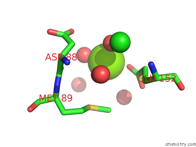
Mono view
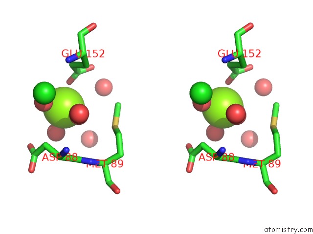
Stereo pair view
Mono view
Stereo pair view
A full contact list of Magnesium with other atoms in the Mg binding
site number 1 of Crystal Structure of Coxsackievirus A24V Soaked with Disialyllacto-N- Tetraose (Dslnt) within 5.0Å range:
|
Reference:
G.Zocher,
N.Mistry,
M.Frank,
I.Hahnlein-Schick,
J.O.Ekstrom,
N.Arnberg,
T.Stehle.
A Sialic Acid Binding Site in A Human Picornavirus. Plos Pathog. V. 10 04401 2014.
ISSN: ISSN 1553-7366
PubMed: 25329320
DOI: 10.1371/JOURNAL.PPAT.1004401
Page generated: Tue Aug 20 01:46:54 2024
ISSN: ISSN 1553-7366
PubMed: 25329320
DOI: 10.1371/JOURNAL.PPAT.1004401
Last articles
Zn in 9J0NZn in 9J0O
Zn in 9J0P
Zn in 9FJX
Zn in 9EKB
Zn in 9C0F
Zn in 9CAH
Zn in 9CH0
Zn in 9CH3
Zn in 9CH1