Magnesium »
PDB 4pyl-4q8b »
4q6x »
Magnesium in PDB 4q6x: Structure of Phospholipase D BETA1B1I From Sicarius Terrosus Venom at 2.14 A Resolution
Enzymatic activity of Structure of Phospholipase D BETA1B1I From Sicarius Terrosus Venom at 2.14 A Resolution
All present enzymatic activity of Structure of Phospholipase D BETA1B1I From Sicarius Terrosus Venom at 2.14 A Resolution:
4.6.1.17;
4.6.1.17;
Protein crystallography data
The structure of Structure of Phospholipase D BETA1B1I From Sicarius Terrosus Venom at 2.14 A Resolution, PDB code: 4q6x
was solved by
D.M.Lajoie,
S.A.Roberts,
P.A.Zobel-Thropp,
G.J.Binford,
M.H.Cordes,
with X-Ray Crystallography technique. A brief refinement statistics is given in the table below:
| Resolution Low / High (Å) | 38.56 / 2.14 |
| Space group | P 32 |
| Cell size a, b, c (Å), α, β, γ (°) | 49.222, 49.222, 90.114, 90.00, 90.00, 120.00 |
| R / Rfree (%) | 16.4 / 22.1 |
Magnesium Binding Sites:
The binding sites of Magnesium atom in the Structure of Phospholipase D BETA1B1I From Sicarius Terrosus Venom at 2.14 A Resolution
(pdb code 4q6x). This binding sites where shown within
5.0 Angstroms radius around Magnesium atom.
In total only one binding site of Magnesium was determined in the Structure of Phospholipase D BETA1B1I From Sicarius Terrosus Venom at 2.14 A Resolution, PDB code: 4q6x:
In total only one binding site of Magnesium was determined in the Structure of Phospholipase D BETA1B1I From Sicarius Terrosus Venom at 2.14 A Resolution, PDB code: 4q6x:
Magnesium binding site 1 out of 1 in 4q6x
Go back to
Magnesium binding site 1 out
of 1 in the Structure of Phospholipase D BETA1B1I From Sicarius Terrosus Venom at 2.14 A Resolution
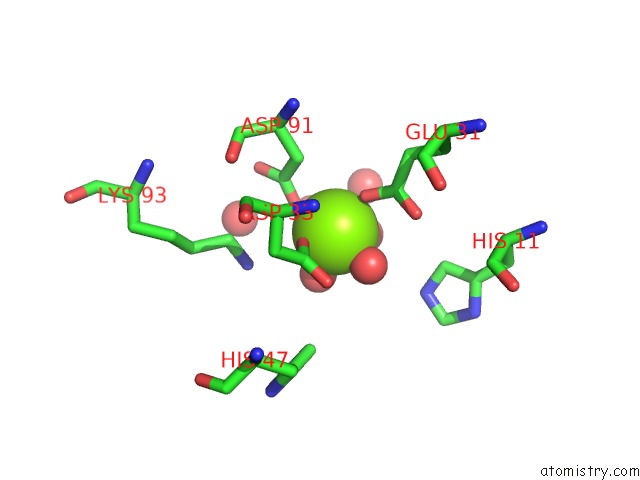
Mono view
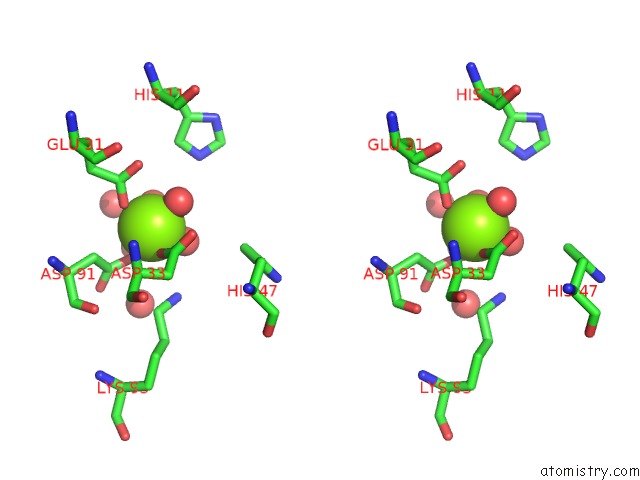
Stereo pair view
Mono view
Stereo pair view
A full contact list of Magnesium with other atoms in the Mg binding
site number 1 of Structure of Phospholipase D BETA1B1I From Sicarius Terrosus Venom at 2.14 A Resolution within 5.0Å range:
|
Reference:
D.M.Lajoie,
S.A.Roberts,
P.A.Zobel-Thropp,
J.L.Delahaye,
V.Bandarian,
G.J.Binford,
M.H.Cordes.
Variable Substrate Preference Among Phospholipase D Toxins From Sicariid Spiders J.Biol.Chem. 2015.
ISSN: ESSN 1083-351X
DOI: 10.1074/JBC.M115.636951
Page generated: Mon Aug 11 22:15:43 2025
ISSN: ESSN 1083-351X
DOI: 10.1074/JBC.M115.636951
Last articles
Mg in 5J32Mg in 5J2T
Mg in 5J2P
Mg in 5J2Q
Mg in 5J02
Mg in 5J01
Mg in 5J2M
Mg in 5J2N
Mg in 5J2K
Mg in 5J2J