Magnesium »
PDB 4q8b-4qgr »
4q8h »
Magnesium in PDB 4q8h: Structure of the Saccharomyces Cerevisiae PAN2 Pseudoubiquitin- Hydrolase-Rnase Module
Enzymatic activity of Structure of the Saccharomyces Cerevisiae PAN2 Pseudoubiquitin- Hydrolase-Rnase Module
All present enzymatic activity of Structure of the Saccharomyces Cerevisiae PAN2 Pseudoubiquitin- Hydrolase-Rnase Module:
3.1.13.4;
3.1.13.4;
Protein crystallography data
The structure of Structure of the Saccharomyces Cerevisiae PAN2 Pseudoubiquitin- Hydrolase-Rnase Module, PDB code: 4q8h
was solved by
I.B.Schaefer,
E.Conti,
with X-Ray Crystallography technique. A brief refinement statistics is given in the table below:
| Resolution Low / High (Å) | 48.01 / 3.10 |
| Space group | I 2 2 2 |
| Cell size a, b, c (Å), α, β, γ (°) | 91.712, 115.505, 257.449, 90.00, 90.00, 90.00 |
| R / Rfree (%) | 23.9 / 27.5 |
Magnesium Binding Sites:
The binding sites of Magnesium atom in the Structure of the Saccharomyces Cerevisiae PAN2 Pseudoubiquitin- Hydrolase-Rnase Module
(pdb code 4q8h). This binding sites where shown within
5.0 Angstroms radius around Magnesium atom.
In total 2 binding sites of Magnesium where determined in the Structure of the Saccharomyces Cerevisiae PAN2 Pseudoubiquitin- Hydrolase-Rnase Module, PDB code: 4q8h:
Jump to Magnesium binding site number: 1; 2;
In total 2 binding sites of Magnesium where determined in the Structure of the Saccharomyces Cerevisiae PAN2 Pseudoubiquitin- Hydrolase-Rnase Module, PDB code: 4q8h:
Jump to Magnesium binding site number: 1; 2;
Magnesium binding site 1 out of 2 in 4q8h
Go back to
Magnesium binding site 1 out
of 2 in the Structure of the Saccharomyces Cerevisiae PAN2 Pseudoubiquitin- Hydrolase-Rnase Module
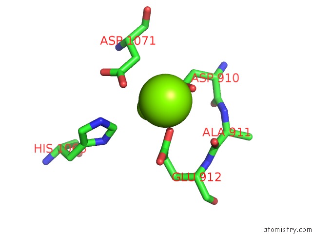
Mono view
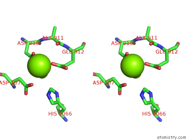
Stereo pair view
Mono view
Stereo pair view
A full contact list of Magnesium with other atoms in the Mg binding
site number 1 of Structure of the Saccharomyces Cerevisiae PAN2 Pseudoubiquitin- Hydrolase-Rnase Module within 5.0Å range:
|
Magnesium binding site 2 out of 2 in 4q8h
Go back to
Magnesium binding site 2 out
of 2 in the Structure of the Saccharomyces Cerevisiae PAN2 Pseudoubiquitin- Hydrolase-Rnase Module
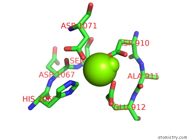
Mono view
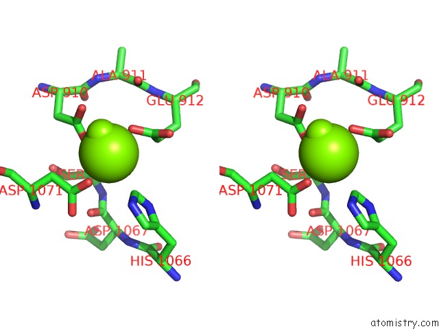
Stereo pair view
Mono view
Stereo pair view
A full contact list of Magnesium with other atoms in the Mg binding
site number 2 of Structure of the Saccharomyces Cerevisiae PAN2 Pseudoubiquitin- Hydrolase-Rnase Module within 5.0Å range:
|
Reference:
I.B.Schafer,
M.Rode,
F.Bonneau,
S.Schussler,
E.Conti.
The Structure of the PAN2-PAN3 Core Complex Reveals Cross-Talk Between Deadenylase and Pseudokinase. Nat.Struct.Mol.Biol. V. 21 591 2014.
ISSN: ISSN 1545-9993
PubMed: 24880344
DOI: 10.1038/NSMB.2834
Page generated: Tue Aug 20 01:51:14 2024
ISSN: ISSN 1545-9993
PubMed: 24880344
DOI: 10.1038/NSMB.2834
Last articles
Zn in 9JYWZn in 9IR4
Zn in 9IR3
Zn in 9GMX
Zn in 9GMW
Zn in 9JEJ
Zn in 9ERF
Zn in 9ERE
Zn in 9EGV
Zn in 9EGW