Magnesium »
PDB 4q8e-4qh0 »
4qa5 »
Magnesium in PDB 4qa5: Crystal Structure of A188T/Y306F HDAC8 in Complex with A Tetrapeptide Substrate
Enzymatic activity of Crystal Structure of A188T/Y306F HDAC8 in Complex with A Tetrapeptide Substrate
All present enzymatic activity of Crystal Structure of A188T/Y306F HDAC8 in Complex with A Tetrapeptide Substrate:
3.5.1.98;
3.5.1.98;
Protein crystallography data
The structure of Crystal Structure of A188T/Y306F HDAC8 in Complex with A Tetrapeptide Substrate, PDB code: 4qa5
was solved by
C.Decroos,
C.B.Bowman,
J.-A.S.Moser,
K.E.Christianson,
M.A.Deardorff,
D.W.Christianson,
with X-Ray Crystallography technique. A brief refinement statistics is given in the table below:
| Resolution Low / High (Å) | 46.42 / 1.76 |
| Space group | P 21 21 21 |
| Cell size a, b, c (Å), α, β, γ (°) | 82.830, 97.760, 105.485, 90.00, 90.00, 90.00 |
| R / Rfree (%) | 16.7 / 19.4 |
Other elements in 4qa5:
The structure of Crystal Structure of A188T/Y306F HDAC8 in Complex with A Tetrapeptide Substrate also contains other interesting chemical elements:
| Potassium | (K) | 4 atoms |
| Zinc | (Zn) | 2 atoms |
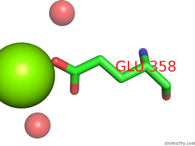
Magnesium Binding Sites:
The binding sites of Magnesium atom in the Crystal Structure of A188T/Y306F HDAC8 in Complex with A Tetrapeptide Substrate
(pdb code 4qa5). This binding sites where shown within
5.0 Angstroms radius around Magnesium atom.
In total only one binding site of Magnesium was determined in the Crystal Structure of A188T/Y306F HDAC8 in Complex with A Tetrapeptide Substrate, PDB code: 4qa5:
In total only one binding site of Magnesium was determined in the Crystal Structure of A188T/Y306F HDAC8 in Complex with A Tetrapeptide Substrate, PDB code: 4qa5:
Magnesium binding site 1 out of 1 in 4qa5
Go back to
Magnesium binding site 1 out
of 1 in the Crystal Structure of A188T/Y306F HDAC8 in Complex with A Tetrapeptide Substrate
Mono view
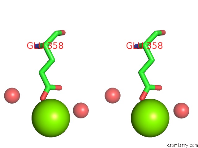
Stereo pair view
Mono view
Stereo pair view
A full contact list of Magnesium with other atoms in the Mg binding
site number 1 of Crystal Structure of A188T/Y306F HDAC8 in Complex with A Tetrapeptide Substrate within 5.0Å range:
|
Reference:
C.Decroos,
C.M.Bowman,
J.A.Moser,
K.E.Christianson,
M.A.Deardorff,
D.W.Christianson.
Compromised Structure and Function of HDAC8 Mutants Identified in Cornelia De Lange Syndrome Spectrum Disorders. Acs Chem.Biol. V. 9 2157 2014.
ISSN: ISSN 1554-8929
PubMed: 25075551
DOI: 10.1021/CB5003762
Page generated: Tue Aug 20 01:51:50 2024
ISSN: ISSN 1554-8929
PubMed: 25075551
DOI: 10.1021/CB5003762
Last articles
Zn in 9MJ5Zn in 9HNW
Zn in 9G0L
Zn in 9FNE
Zn in 9DZN
Zn in 9E0I
Zn in 9D32
Zn in 9DAK
Zn in 8ZXC
Zn in 8ZUF