Magnesium »
PDB 4qh0-4qpm »
4qh0 »
Magnesium in PDB 4qh0: Crystal Structure of Nuca From Streptococcus Agalactiae with Magnesium Ion Bound
Protein crystallography data
The structure of Crystal Structure of Nuca From Streptococcus Agalactiae with Magnesium Ion Bound, PDB code: 4qh0
was solved by
A.F.Moon,
P.Gaudu,
L.C.Pedersen,
with X-Ray Crystallography technique. A brief refinement statistics is given in the table below:
| Resolution Low / High (Å) | 24.85 / 2.00 |
| Space group | P 63 |
| Cell size a, b, c (Å), α, β, γ (°) | 123.721, 123.721, 157.403, 90.00, 90.00, 120.00 |
| R / Rfree (%) | 15.5 / 19 |
Other elements in 4qh0:
The structure of Crystal Structure of Nuca From Streptococcus Agalactiae with Magnesium Ion Bound also contains other interesting chemical elements:
| Chlorine | (Cl) | 6 atoms |
Magnesium Binding Sites:
The binding sites of Magnesium atom in the Crystal Structure of Nuca From Streptococcus Agalactiae with Magnesium Ion Bound
(pdb code 4qh0). This binding sites where shown within
5.0 Angstroms radius around Magnesium atom.
In total 4 binding sites of Magnesium where determined in the Crystal Structure of Nuca From Streptococcus Agalactiae with Magnesium Ion Bound, PDB code: 4qh0:
Jump to Magnesium binding site number: 1; 2; 3; 4;
In total 4 binding sites of Magnesium where determined in the Crystal Structure of Nuca From Streptococcus Agalactiae with Magnesium Ion Bound, PDB code: 4qh0:
Jump to Magnesium binding site number: 1; 2; 3; 4;
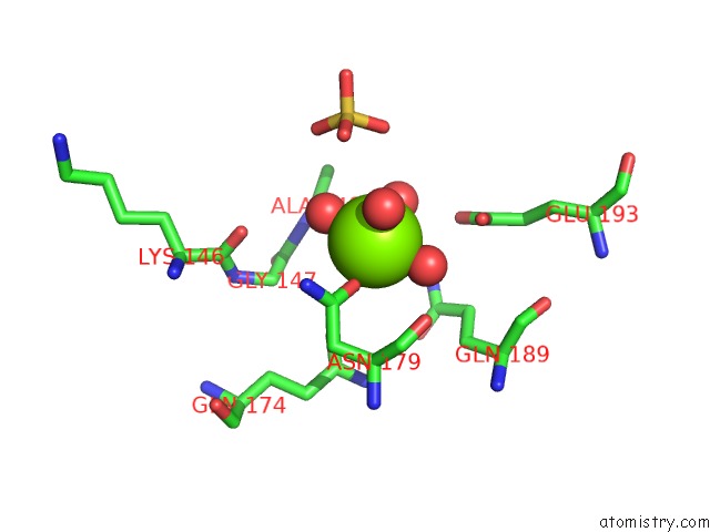
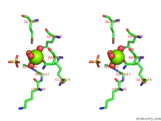
Magnesium binding site 1 out of 4 in 4qh0
Go back to
Magnesium binding site 1 out
of 4 in the Crystal Structure of Nuca From Streptococcus Agalactiae with Magnesium Ion Bound
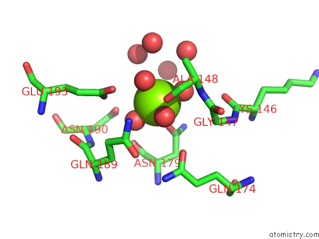
Mono view
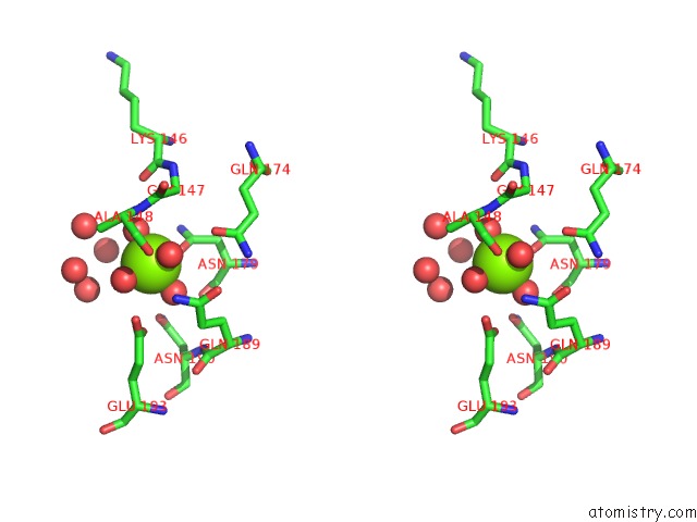
Stereo pair view
Mono view
Stereo pair view
A full contact list of Magnesium with other atoms in the Mg binding
site number 1 of Crystal Structure of Nuca From Streptococcus Agalactiae with Magnesium Ion Bound within 5.0Å range:
|
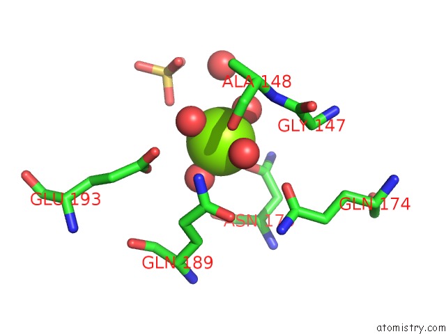
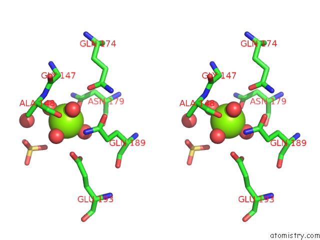
Magnesium binding site 2 out of 4 in 4qh0
Go back to
Magnesium binding site 2 out
of 4 in the Crystal Structure of Nuca From Streptococcus Agalactiae with Magnesium Ion Bound
Mono view
Stereo pair view
Mono view
Stereo pair view
A full contact list of Magnesium with other atoms in the Mg binding
site number 2 of Crystal Structure of Nuca From Streptococcus Agalactiae with Magnesium Ion Bound within 5.0Å range:
|
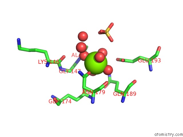
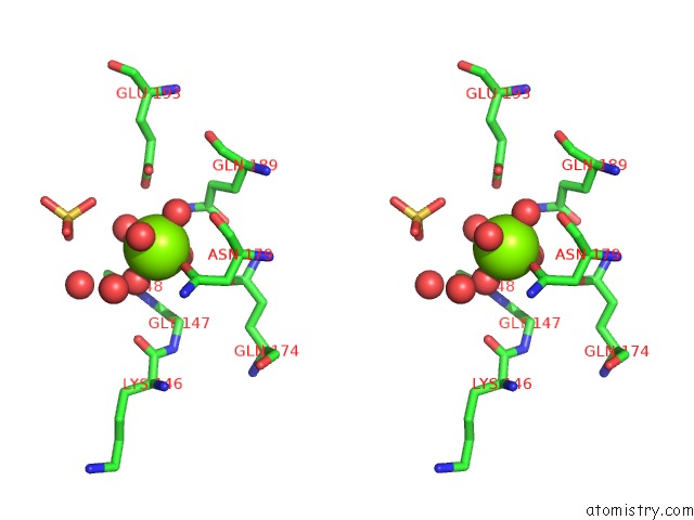
Magnesium binding site 3 out of 4 in 4qh0
Go back to
Magnesium binding site 3 out
of 4 in the Crystal Structure of Nuca From Streptococcus Agalactiae with Magnesium Ion Bound
Mono view
Stereo pair view
Mono view
Stereo pair view
A full contact list of Magnesium with other atoms in the Mg binding
site number 3 of Crystal Structure of Nuca From Streptococcus Agalactiae with Magnesium Ion Bound within 5.0Å range:
|
Magnesium binding site 4 out of 4 in 4qh0
Go back to
Magnesium binding site 4 out
of 4 in the Crystal Structure of Nuca From Streptococcus Agalactiae with Magnesium Ion Bound
Mono view
Stereo pair view
Mono view
Stereo pair view
A full contact list of Magnesium with other atoms in the Mg binding
site number 4 of Crystal Structure of Nuca From Streptococcus Agalactiae with Magnesium Ion Bound within 5.0Å range:
|
Reference:
A.F.Moon,
P.Gaudu,
L.C.Pedersen.
Structural Characterization of the Virulence Factor Nuclease A From Streptococcus Agalactiae. Acta Crystallogr.,Sect.D V. 70 2937 2014.
ISSN: ISSN 0907-4449
PubMed: 25372684
DOI: 10.1107/S1399004714019725
Page generated: Tue Aug 20 01:59:05 2024
ISSN: ISSN 0907-4449
PubMed: 25372684
DOI: 10.1107/S1399004714019725
Last articles
Zn in 9J0NZn in 9J0O
Zn in 9J0P
Zn in 9FJX
Zn in 9EKB
Zn in 9C0F
Zn in 9CAH
Zn in 9CH0
Zn in 9CH3
Zn in 9CH1