Magnesium »
PDB 4qhe-4qpz »
4qhz »
Magnesium in PDB 4qhz: Crystal Structure of A Putative Glycosyl Hydrolase (BDI_3914) From Parabacteroides Distasonis Atcc 8503 at 2.13 A Resolution
Protein crystallography data
The structure of Crystal Structure of A Putative Glycosyl Hydrolase (BDI_3914) From Parabacteroides Distasonis Atcc 8503 at 2.13 A Resolution, PDB code: 4qhz
was solved by
Joint Center For Structural Genomics (Jcsg),
with X-Ray Crystallography technique. A brief refinement statistics is given in the table below:
| Resolution Low / High (Å) | 48.25 / 2.13 |
| Space group | P 1 21 1 |
| Cell size a, b, c (Å), α, β, γ (°) | 47.774, 144.763, 82.452, 90.00, 101.27, 90.00 |
| R / Rfree (%) | 17.1 / 20.3 |
Magnesium Binding Sites:
The binding sites of Magnesium atom in the Crystal Structure of A Putative Glycosyl Hydrolase (BDI_3914) From Parabacteroides Distasonis Atcc 8503 at 2.13 A Resolution
(pdb code 4qhz). This binding sites where shown within
5.0 Angstroms radius around Magnesium atom.
In total 4 binding sites of Magnesium where determined in the Crystal Structure of A Putative Glycosyl Hydrolase (BDI_3914) From Parabacteroides Distasonis Atcc 8503 at 2.13 A Resolution, PDB code: 4qhz:
Jump to Magnesium binding site number: 1; 2; 3; 4;
In total 4 binding sites of Magnesium where determined in the Crystal Structure of A Putative Glycosyl Hydrolase (BDI_3914) From Parabacteroides Distasonis Atcc 8503 at 2.13 A Resolution, PDB code: 4qhz:
Jump to Magnesium binding site number: 1; 2; 3; 4;
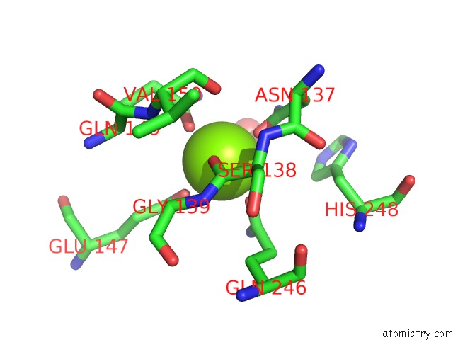
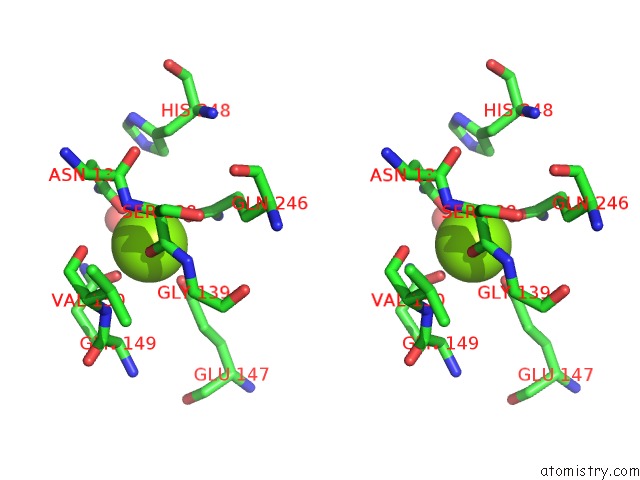
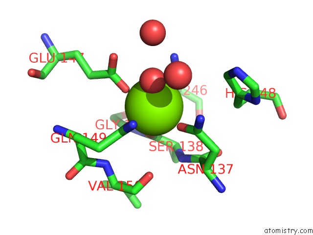
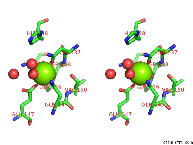
Magnesium binding site 1 out of 4 in 4qhz
Go back to
Magnesium binding site 1 out
of 4 in the Crystal Structure of A Putative Glycosyl Hydrolase (BDI_3914) From Parabacteroides Distasonis Atcc 8503 at 2.13 A Resolution
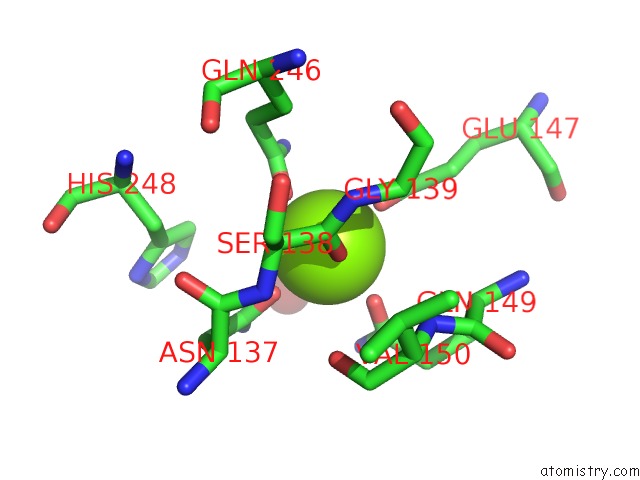
Mono view
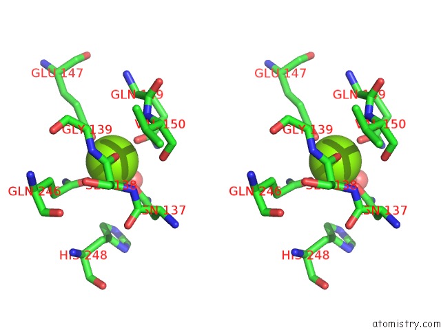
Stereo pair view
Mono view
Stereo pair view
A full contact list of Magnesium with other atoms in the Mg binding
site number 1 of Crystal Structure of A Putative Glycosyl Hydrolase (BDI_3914) From Parabacteroides Distasonis Atcc 8503 at 2.13 A Resolution within 5.0Å range:
|
Magnesium binding site 2 out of 4 in 4qhz
Go back to
Magnesium binding site 2 out
of 4 in the Crystal Structure of A Putative Glycosyl Hydrolase (BDI_3914) From Parabacteroides Distasonis Atcc 8503 at 2.13 A Resolution
Mono view
Stereo pair view
Mono view
Stereo pair view
A full contact list of Magnesium with other atoms in the Mg binding
site number 2 of Crystal Structure of A Putative Glycosyl Hydrolase (BDI_3914) From Parabacteroides Distasonis Atcc 8503 at 2.13 A Resolution within 5.0Å range:
|
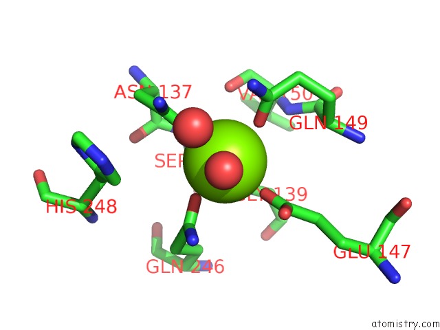
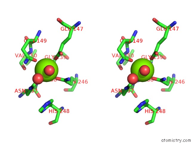
Magnesium binding site 3 out of 4 in 4qhz
Go back to
Magnesium binding site 3 out
of 4 in the Crystal Structure of A Putative Glycosyl Hydrolase (BDI_3914) From Parabacteroides Distasonis Atcc 8503 at 2.13 A Resolution
Mono view
Stereo pair view
Mono view
Stereo pair view
A full contact list of Magnesium with other atoms in the Mg binding
site number 3 of Crystal Structure of A Putative Glycosyl Hydrolase (BDI_3914) From Parabacteroides Distasonis Atcc 8503 at 2.13 A Resolution within 5.0Å range:
|
Magnesium binding site 4 out of 4 in 4qhz
Go back to
Magnesium binding site 4 out
of 4 in the Crystal Structure of A Putative Glycosyl Hydrolase (BDI_3914) From Parabacteroides Distasonis Atcc 8503 at 2.13 A Resolution
Mono view
Stereo pair view
Mono view
Stereo pair view
A full contact list of Magnesium with other atoms in the Mg binding
site number 4 of Crystal Structure of A Putative Glycosyl Hydrolase (BDI_3914) From Parabacteroides Distasonis Atcc 8503 at 2.13 A Resolution within 5.0Å range:
|
Reference:
Joint Center For Structural Genomics (Jcsg),
Joint Center For Structural Genomics (Jcsg).
N/A N/A.
Page generated: Mon Aug 11 22:25:00 2025
Last articles
Mg in 5J04Mg in 5IYZ
Mg in 5IZL
Mg in 5IYS
Mg in 5IYD
Mg in 5IYC
Mg in 5IYB
Mg in 5IY9
Mg in 5IYA
Mg in 5IY8