Magnesium »
PDB 4qh0-4qpm »
4ql3 »
Magnesium in PDB 4ql3: Crystal Structure of A Gdp-Bound G12R Oncogenic Mutant of Human Gtpase Kras
Enzymatic activity of Crystal Structure of A Gdp-Bound G12R Oncogenic Mutant of Human Gtpase Kras
All present enzymatic activity of Crystal Structure of A Gdp-Bound G12R Oncogenic Mutant of Human Gtpase Kras:
3.6.5.2;
3.6.5.2;
Protein crystallography data
The structure of Crystal Structure of A Gdp-Bound G12R Oncogenic Mutant of Human Gtpase Kras, PDB code: 4ql3
was solved by
J.C.Hunter,
A.Manandhar,
D.Gurbani,
Z.Chen,
K.D.Westover,
with X-Ray Crystallography technique. A brief refinement statistics is given in the table below:
| Resolution Low / High (Å) | 24.50 / 1.04 |
| Space group | P 21 21 21 |
| Cell size a, b, c (Å), α, β, γ (°) | 39.125, 40.908, 91.806, 90.00, 90.00, 90.00 |
| R / Rfree (%) | 13.8 / 16.2 |
Magnesium Binding Sites:
The binding sites of Magnesium atom in the Crystal Structure of A Gdp-Bound G12R Oncogenic Mutant of Human Gtpase Kras
(pdb code 4ql3). This binding sites where shown within
5.0 Angstroms radius around Magnesium atom.
In total only one binding site of Magnesium was determined in the Crystal Structure of A Gdp-Bound G12R Oncogenic Mutant of Human Gtpase Kras, PDB code: 4ql3:
In total only one binding site of Magnesium was determined in the Crystal Structure of A Gdp-Bound G12R Oncogenic Mutant of Human Gtpase Kras, PDB code: 4ql3:
Magnesium binding site 1 out of 1 in 4ql3
Go back to
Magnesium binding site 1 out
of 1 in the Crystal Structure of A Gdp-Bound G12R Oncogenic Mutant of Human Gtpase Kras
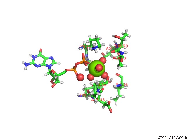
Mono view
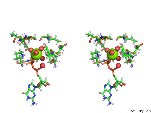
Stereo pair view
Mono view
Stereo pair view
A full contact list of Magnesium with other atoms in the Mg binding
site number 1 of Crystal Structure of A Gdp-Bound G12R Oncogenic Mutant of Human Gtpase Kras within 5.0Å range:
|
Reference:
J.C.Hunter,
A.Manandhar,
M.A.Carrasco,
D.Gurbani,
S.Gondi,
K.D.Westover.
Biochemical and Structural Analysis of Common Cancer-Associated Kras Mutations. Mol Cancer Res. V. 13 1325 2015.
ISSN: ISSN 1541-7786
PubMed: 26037647
DOI: 10.1158/1541-7786.MCR-15-0203
Page generated: Tue Aug 20 02:02:22 2024
ISSN: ISSN 1541-7786
PubMed: 26037647
DOI: 10.1158/1541-7786.MCR-15-0203
Last articles
Zn in 9J0NZn in 9J0O
Zn in 9J0P
Zn in 9FJX
Zn in 9EKB
Zn in 9C0F
Zn in 9CAH
Zn in 9CH0
Zn in 9CH3
Zn in 9CH1