Atomistry » Magnesium » PDB 4qq8-4qwi » 4qq8
Atomistry »
  Magnesium »
    PDB 4qq8-4qwi »
      4qq8 »

Magnesium in PDB 4qq8: Crystal Structure of the Formolase Fls in Space Group P 43 21 2

Protein crystallography data

The structure of Crystal Structure of the Formolase Fls in Space Group P 43 21 2, PDB code: 4qq8 was solved by B.W.Shen, J.B.Siegel, B.L.Stoddard, with X-Ray Crystallography technique. A brief refinement statistics is given in the table below:

Resolution Low / High (Å) 127.52 / 2.88
Space group P 43 21 2
Cell size a, b, c (Å), α, β, γ (°) 144.742, 144.742, 269.553, 90.00, 90.00, 90.00
R / Rfree (%) 16.9 / 19.9

Magnesium Binding Sites:

The binding sites of Magnesium atom in the Crystal Structure of the Formolase Fls in Space Group P 43 21 2 (pdb code 4qq8). This binding sites where shown within 5.0 Angstroms radius around Magnesium atom.
In total 4 binding sites of Magnesium where determined in the Crystal Structure of the Formolase Fls in Space Group P 43 21 2, PDB code: 4qq8:
Jump to Magnesium binding site number: 1; 2; 3; 4;

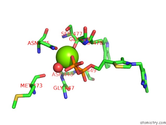
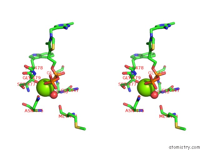
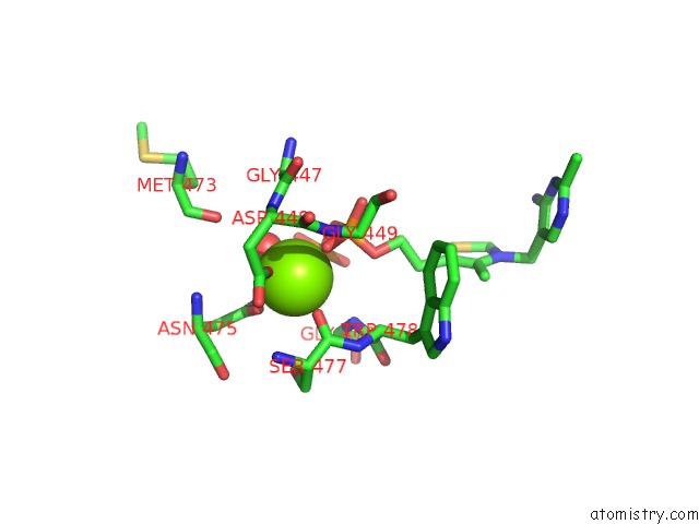
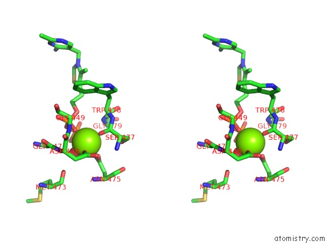
Magnesium binding site 1 out of 4 in 4qq8

Go back to Magnesium Binding Sites List in 4qq8
Magnesium binding site 1 out of 4 in the Crystal Structure of the Formolase Fls in Space Group P 43 21 2


Mono view


Stereo pair view

A full contact list of Magnesium with other atoms in the Mg binding site number 1 of Crystal Structure of the Formolase Fls in Space Group P 43 21 2 within 5.0Å range:
probe atom residue distance (Å) B Occ
A:Mg601

b:39.0
occ:1.00
O1A A:TPP602 1.9 45.9 1.0
O A:SER477 2.0 47.5 1.0
O1B A:TPP602 2.0 43.5 1.0
O A:HOH703 2.1 33.0 1.0
OD1 A:ASN475 2.2 42.8 1.0
OD1 A:ASP448 2.2 46.1 1.0
CG A:ASN475 3.1 44.5 1.0
PA A:TPP602 3.1 44.1 1.0
PB A:TPP602 3.2 44.2 1.0
C A:SER477 3.2 46.3 1.0
ND2 A:ASN475 3.4 45.8 1.0
O3A A:TPP602 3.4 44.7 1.0
CG A:ASP448 3.4 47.6 1.0
N A:GLY479 3.7 50.0 1.0
O2B A:TPP602 3.8 44.0 1.0
O7 A:TPP602 3.8 46.6 1.0
N A:ASP448 4.0 49.3 1.0
N A:SER477 4.0 46.5 1.0
OD2 A:ASP448 4.0 48.3 1.0
CA A:SER477 4.1 46.9 1.0
OG A:SER477 4.1 48.0 1.0
N A:TRP478 4.2 45.9 1.0
N A:GLY449 4.2 46.0 1.0
CA A:GLY479 4.3 51.5 1.0
CA A:TRP478 4.4 46.6 1.0
O3B A:TPP602 4.4 43.0 1.0
O2A A:TPP602 4.4 40.9 1.0
CB A:ASN475 4.5 45.7 1.0
N A:ASN475 4.5 46.6 1.0
O A:MET473 4.5 45.9 1.0
C A:TRP478 4.5 48.5 1.0
CB A:ASP448 4.6 49.1 1.0
CA A:ASP448 4.6 48.9 1.0
CA A:GLY447 4.7 48.0 1.0
C A:GLY447 4.7 48.6 1.0
CB A:SER477 4.8 48.2 1.0
CA A:ASN475 4.9 47.1 1.0
C A:ASN475 4.9 48.8 1.0
C A:ASP448 4.9 47.6 1.0

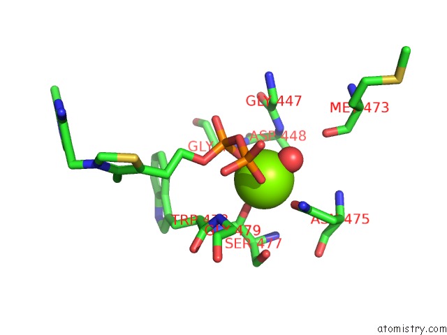
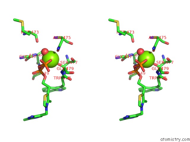
Magnesium binding site 2 out of 4 in 4qq8

Go back to Magnesium Binding Sites List in 4qq8
Magnesium binding site 2 out of 4 in the Crystal Structure of the Formolase Fls in Space Group P 43 21 2


Mono view


Stereo pair view

A full contact list of Magnesium with other atoms in the Mg binding site number 2 of Crystal Structure of the Formolase Fls in Space Group P 43 21 2 within 5.0Å range:
probe atom residue distance (Å) B Occ
B:Mg601

b:34.7
occ:1.00
O1A B:TPP602 2.0 48.2 1.0
O3B B:TPP602 2.0 46.8 1.0
OD1 B:ASP448 2.1 48.0 1.0
OD1 B:ASN475 2.1 52.7 1.0
O B:SER477 2.6 45.6 1.0
CG B:ASN475 3.0 52.9 1.0
PB B:TPP602 3.1 47.2 1.0
O1B B:TPP602 3.2 48.0 1.0
ND2 B:ASN475 3.3 53.6 1.0
PA B:TPP602 3.3 46.2 1.0
CG B:ASP448 3.3 48.7 1.0
O3A B:TPP602 3.6 46.6 1.0
N B:ASP448 3.6 47.5 1.0
O B:MET473 3.8 54.8 1.0
C B:SER477 3.8 47.9 1.0
N B:ASN475 4.1 52.1 1.0
OD2 B:ASP448 4.1 50.6 1.0
CA B:GLY447 4.3 46.0 1.0
O7 B:TPP602 4.3 49.6 1.0
O2A B:TPP602 4.3 43.9 1.0
N B:GLY479 4.3 50.7 1.0
N B:GLY449 4.3 48.0 1.0
CB B:ASN475 4.3 52.5 1.0
CB B:ASP448 4.4 48.2 1.0
C B:GLY447 4.4 46.2 1.0
O2B B:TPP602 4.4 46.0 1.0
N B:SER477 4.4 51.5 1.0
CA B:ASP448 4.4 47.2 1.0
OG B:SER477 4.6 53.9 1.0
CA B:ASN475 4.6 53.3 1.0
CA B:SER477 4.6 50.5 1.0
CA B:GLY479 4.7 49.4 1.0
N B:TRP478 4.8 47.3 1.0
OG1 B:THR396 4.9 56.4 1.0
C B:ASN475 4.9 54.0 1.0
C B:ASP448 4.9 47.0 1.0
C B:MET473 5.0 54.4 1.0

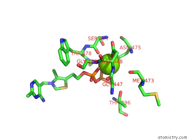
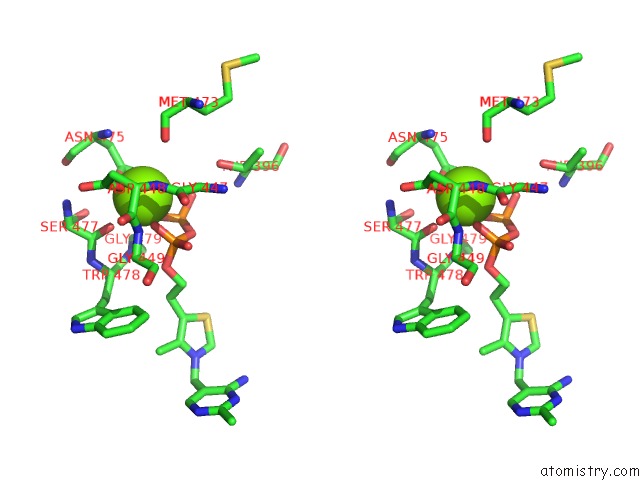
Magnesium binding site 3 out of 4 in 4qq8

Go back to Magnesium Binding Sites List in 4qq8
Magnesium binding site 3 out of 4 in the Crystal Structure of the Formolase Fls in Space Group P 43 21 2


Mono view


Stereo pair view

A full contact list of Magnesium with other atoms in the Mg binding site number 3 of Crystal Structure of the Formolase Fls in Space Group P 43 21 2 within 5.0Å range:
probe atom residue distance (Å) B Occ
C:Mg601

b:33.4
occ:1.00
OD1 C:ASP448 2.0 39.5 1.0
O C:SER477 2.0 39.6 1.0
O1A C:TPP602 2.0 38.2 1.0
O A:HOH704 2.0 28.5 1.0
O3B C:TPP602 2.2 40.5 1.0
OD1 C:ASN475 2.2 38.5 1.0
CG C:ASP448 3.2 40.6 1.0
CG C:ASN475 3.2 38.5 1.0
PA C:TPP602 3.2 37.1 1.0
C C:SER477 3.2 39.7 1.0
PB C:TPP602 3.3 40.6 1.0
O3A C:TPP602 3.6 38.9 1.0
ND2 C:ASN475 3.6 37.0 1.0
N C:ASP448 3.7 39.0 1.0
OD2 C:ASP448 3.8 40.2 1.0
O7 C:TPP602 3.8 41.5 1.0
N C:GLY479 3.9 42.6 1.0
N C:SER477 4.0 41.6 1.0
N C:GLY449 4.0 40.7 1.0
O1B C:TPP602 4.0 38.8 1.0
CA C:SER477 4.1 40.0 1.0
N C:TRP478 4.2 40.8 1.0
OG C:SER477 4.3 37.8 1.0
CB C:ASP448 4.3 41.0 1.0
CA C:ASP448 4.4 39.3 1.0
O C:MET473 4.4 49.8 1.0
N C:ASN475 4.4 44.8 1.0
CA C:TRP478 4.4 41.4 1.0
O2A C:TPP602 4.5 35.1 1.0
C C:GLY447 4.5 39.4 1.0
CA C:GLY479 4.5 42.0 1.0
O2B C:TPP602 4.5 40.9 1.0
CB C:ASN475 4.6 40.9 1.0
CA C:GLY447 4.6 38.7 1.0
C C:ASP448 4.6 40.2 1.0
C C:TRP478 4.7 43.0 1.0
CA C:ASN475 4.8 43.0 1.0
CB C:SER477 4.9 39.1 1.0
C C:ASN475 4.9 43.1 1.0
CA C:GLY449 4.9 42.3 1.0

Magnesium binding site 4 out of 4 in 4qq8

Go back to Magnesium Binding Sites List in 4qq8
Magnesium binding site 4 out of 4 in the Crystal Structure of the Formolase Fls in Space Group P 43 21 2


Mono view


Stereo pair view

A full contact list of Magnesium with other atoms in the Mg binding site number 4 of Crystal Structure of the Formolase Fls in Space Group P 43 21 2 within 5.0Å range:
probe atom residue distance (Å) B Occ
D:Mg601

b:37.1
occ:1.00
O1A D:TPP602 1.9 43.5 1.0
O A:HOH705 1.9 40.1 1.0
O D:SER477 2.0 48.1 1.0
OD1 D:ASP448 2.0 48.9 1.0
O3B D:TPP602 2.2 42.5 1.0
OD1 D:ASN475 2.3 48.0 1.0
PA D:TPP602 3.1 42.7 1.0
CG D:ASP448 3.2 52.8 1.0
C D:SER477 3.2 48.0 1.0
PB D:TPP602 3.3 43.0 1.0
CG D:ASN475 3.3 47.7 1.0
O3A D:TPP602 3.3 43.4 1.0
ND2 D:ASN475 3.7 48.9 1.0
O7 D:TPP602 3.7 46.0 1.0
N D:ASP448 3.8 46.9 1.0
N D:GLY479 3.9 47.0 1.0
OD2 D:ASP448 3.9 55.4 1.0
N D:GLY449 4.0 56.9 1.0
N D:SER477 4.0 51.4 1.0
CA D:SER477 4.1 50.8 1.0
N D:TRP478 4.2 46.2 1.0
O2B D:TPP602 4.2 42.4 1.0
OG D:SER477 4.3 50.3 1.0
CB D:ASP448 4.3 51.5 1.0
CA D:TRP478 4.4 47.2 1.0
O2A D:TPP602 4.4 40.7 1.0
CA D:ASP448 4.4 50.1 1.0
O1B D:TPP602 4.4 42.4 1.0
O D:MET473 4.5 52.5 1.0
N D:ASN475 4.5 49.6 1.0
CA D:GLY479 4.5 48.5 1.0
C D:GLY447 4.5 44.9 1.0
CA D:GLY447 4.6 44.2 1.0
C D:TRP478 4.6 46.6 1.0
C D:ASP448 4.6 53.2 1.0
CB D:ASN475 4.7 49.4 1.0
CA D:GLY449 4.9 55.6 1.0
CB D:SER477 4.9 51.1 1.0
CA D:ASN475 4.9 50.2 1.0
C D:ASN475 4.9 50.6 1.0

Reference:

J.B.Siegel, A.L Smith, S.Poust, A.Wargacki, A.Bar-Even, C.Louw, B.W.Shen, C.B.Eiben, H.Tran, E.Noor. Computational Protein Design Enables A Novel One-Carbon Assimilation Pathway To Be Published.
Page generated: Tue Aug 20 02:08:40 2024

Last articles

Zn in 9MJ5
Zn in 9HNW
Zn in 9G0L
Zn in 9FNE
Zn in 9DZN
Zn in 9E0I
Zn in 9D32
Zn in 9DAK
Zn in 8ZXC
Zn in 8ZUF
© Copyright 2008-2020 by atomistry.com
Home   |    Site Map   |    Copyright   |    Contact us   |    Privacy