Magnesium »
PDB 4r17-4rcy »
4r82 »
Magnesium in PDB 4r82: The Crystal Structure of An Oxidoreductase (SGCE6)From Streptomyces Globisporus in Complex with Fad and Nad
Protein crystallography data
The structure of The Crystal Structure of An Oxidoreductase (SGCE6)From Streptomyces Globisporus in Complex with Fad and Nad, PDB code: 4r82
was solved by
K.Tan,
L.Bigelow,
S.Clancy,
G.Babnigg,
C.A.Bingman,
R.Yennamalli,
J.Lohman,
M.Ma,
B.Shen,
G.N.Phillips Jr.,
A.Joachimiak,
Midwest Center Forstructural Genomics (Mcsg),
Enzyme Discovery For Natural Productbiosynthesis (Natpro),
with X-Ray Crystallography technique. A brief refinement statistics is given in the table below:
| Resolution Low / High (Å) | 34.93 / 1.66 |
| Space group | P 1 21 1 |
| Cell size a, b, c (Å), α, β, γ (°) | 42.046, 62.830, 70.089, 90.00, 92.06, 90.00 |
| R / Rfree (%) | 16.3 / 18.8 |
Other elements in 4r82:
The structure of The Crystal Structure of An Oxidoreductase (SGCE6)From Streptomyces Globisporus in Complex with Fad and Nad also contains other interesting chemical elements:
| Calcium | (Ca) | 3 atoms |
| Chlorine | (Cl) | 2 atoms |
Magnesium Binding Sites:
The binding sites of Magnesium atom in the The Crystal Structure of An Oxidoreductase (SGCE6)From Streptomyces Globisporus in Complex with Fad and Nad
(pdb code 4r82). This binding sites where shown within
5.0 Angstroms radius around Magnesium atom.
In total only one binding site of Magnesium was determined in the The Crystal Structure of An Oxidoreductase (SGCE6)From Streptomyces Globisporus in Complex with Fad and Nad, PDB code: 4r82:
In total only one binding site of Magnesium was determined in the The Crystal Structure of An Oxidoreductase (SGCE6)From Streptomyces Globisporus in Complex with Fad and Nad, PDB code: 4r82:
Magnesium binding site 1 out of 1 in 4r82
Go back to
Magnesium binding site 1 out
of 1 in the The Crystal Structure of An Oxidoreductase (SGCE6)From Streptomyces Globisporus in Complex with Fad and Nad
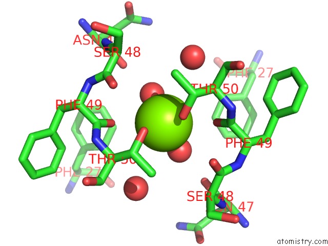
Mono view
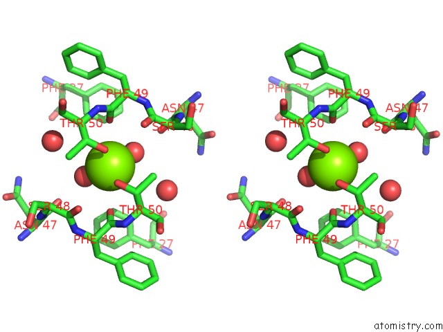
Stereo pair view
Mono view
Stereo pair view
A full contact list of Magnesium with other atoms in the Mg binding
site number 1 of The Crystal Structure of An Oxidoreductase (SGCE6)From Streptomyces Globisporus in Complex with Fad and Nad within 5.0Å range:
|
Reference:
K.Tan,
L.Bigelow,
S.Clancy,
G.Babnigg,
C.A.Bingman,
R.Yennamalli,
J.Lohman,
M.Ma,
B.Shen,
G.N.Phillips Jr.,
A.Joachimiak.
The Crystal Structure of An Oxidoreductase (SGCE6)From Streptomyces Globisporus in Complex with Fad and Nad. To Be Published.
Page generated: Tue Aug 20 02:54:41 2024
Last articles
Fe in 2YXOFe in 2YRS
Fe in 2YXC
Fe in 2YNM
Fe in 2YVJ
Fe in 2YP1
Fe in 2YU2
Fe in 2YU1
Fe in 2YQB
Fe in 2YOO