Magnesium »
PDB 4r17-4rcy »
4r9m »
Magnesium in PDB 4r9m: Crystal Structure of Spermidine N-Acetyltransferase From Escherichia Coli
Enzymatic activity of Crystal Structure of Spermidine N-Acetyltransferase From Escherichia Coli
All present enzymatic activity of Crystal Structure of Spermidine N-Acetyltransferase From Escherichia Coli:
2.3.1.57;
2.3.1.57;
Protein crystallography data
The structure of Crystal Structure of Spermidine N-Acetyltransferase From Escherichia Coli, PDB code: 4r9m
was solved by
E.V.Filippova,
G.Minasov,
O.Kiryukhina,
L.Shuvalova,
S.Grimshaw,
A.J.Wolfe,
W.F.Anderson,
Center For Structural Genomics Of Infectiousdiseases (Csgid),
with X-Ray Crystallography technique. A brief refinement statistics is given in the table below:
| Resolution Low / High (Å) | 29.61 / 2.90 |
| Space group | P 42 21 2 |
| Cell size a, b, c (Å), α, β, γ (°) | 110.919, 110.919, 108.505, 90.00, 90.00, 90.00 |
| R / Rfree (%) | 17.6 / 25.9 |
Magnesium Binding Sites:
The binding sites of Magnesium atom in the Crystal Structure of Spermidine N-Acetyltransferase From Escherichia Coli
(pdb code 4r9m). This binding sites where shown within
5.0 Angstroms radius around Magnesium atom.
In total only one binding site of Magnesium was determined in the Crystal Structure of Spermidine N-Acetyltransferase From Escherichia Coli, PDB code: 4r9m:
In total only one binding site of Magnesium was determined in the Crystal Structure of Spermidine N-Acetyltransferase From Escherichia Coli, PDB code: 4r9m:
Magnesium binding site 1 out of 1 in 4r9m
Go back to
Magnesium binding site 1 out
of 1 in the Crystal Structure of Spermidine N-Acetyltransferase From Escherichia Coli
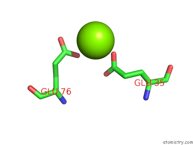
Mono view
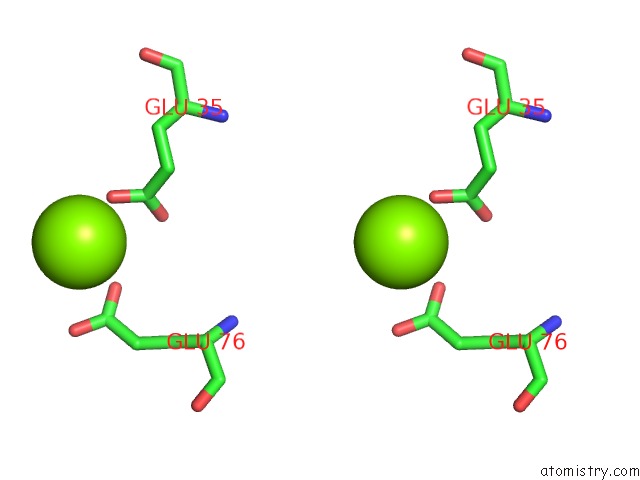
Stereo pair view
Mono view
Stereo pair view
A full contact list of Magnesium with other atoms in the Mg binding
site number 1 of Crystal Structure of Spermidine N-Acetyltransferase From Escherichia Coli within 5.0Å range:
|
Reference:
E.V.Filippova,
G.Minasov,
O.Kiryukhina,
L.Shuvalova,
S.Grimshaw,
A.J.Wolfe,
W.F.Anderson.
Crystal Structure of Spermidine N-Acetyltransferase From Escherichia Coli To Be Published.
Page generated: Tue Aug 20 02:56:21 2024
Last articles
Ca in 2XGQCa in 2XHN
Ca in 2XGP
Ca in 2XJ7
Ca in 2XHI
Ca in 2XHJ
Ca in 2XHH
Ca in 2XF2
Ca in 2XFV
Ca in 2XFG