Magnesium »
PDB 4rcy-4rke »
4rf5 »
Magnesium in PDB 4rf5: Crystal Structure of Ketoreductase From Lactobacillus Kefir, E145S Mutant
Protein crystallography data
The structure of Crystal Structure of Ketoreductase From Lactobacillus Kefir, E145S Mutant, PDB code: 4rf5
was solved by
Y.Tang,
N.Tibrewal,
D.Cascio,
with X-Ray Crystallography technique. A brief refinement statistics is given in the table below:
| Resolution Low / High (Å) | 69.56 / 1.60 |
| Space group | P 21 21 2 |
| Cell size a, b, c (Å), α, β, γ (°) | 66.140, 110.340, 69.560, 90.00, 90.00, 90.00 |
| R / Rfree (%) | 16.3 / 18.3 |
Magnesium Binding Sites:
The binding sites of Magnesium atom in the Crystal Structure of Ketoreductase From Lactobacillus Kefir, E145S Mutant
(pdb code 4rf5). This binding sites where shown within
5.0 Angstroms radius around Magnesium atom.
In total 2 binding sites of Magnesium where determined in the Crystal Structure of Ketoreductase From Lactobacillus Kefir, E145S Mutant, PDB code: 4rf5:
Jump to Magnesium binding site number: 1; 2;
In total 2 binding sites of Magnesium where determined in the Crystal Structure of Ketoreductase From Lactobacillus Kefir, E145S Mutant, PDB code: 4rf5:
Jump to Magnesium binding site number: 1; 2;
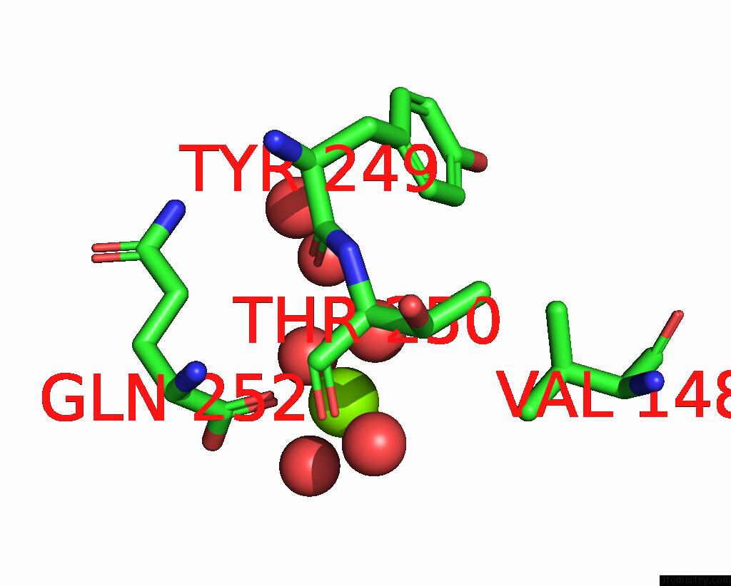
Magnesium binding site 1 out of 2 in 4rf5
Go back to
Magnesium binding site 1 out
of 2 in the Crystal Structure of Ketoreductase From Lactobacillus Kefir, E145S Mutant
Mono view
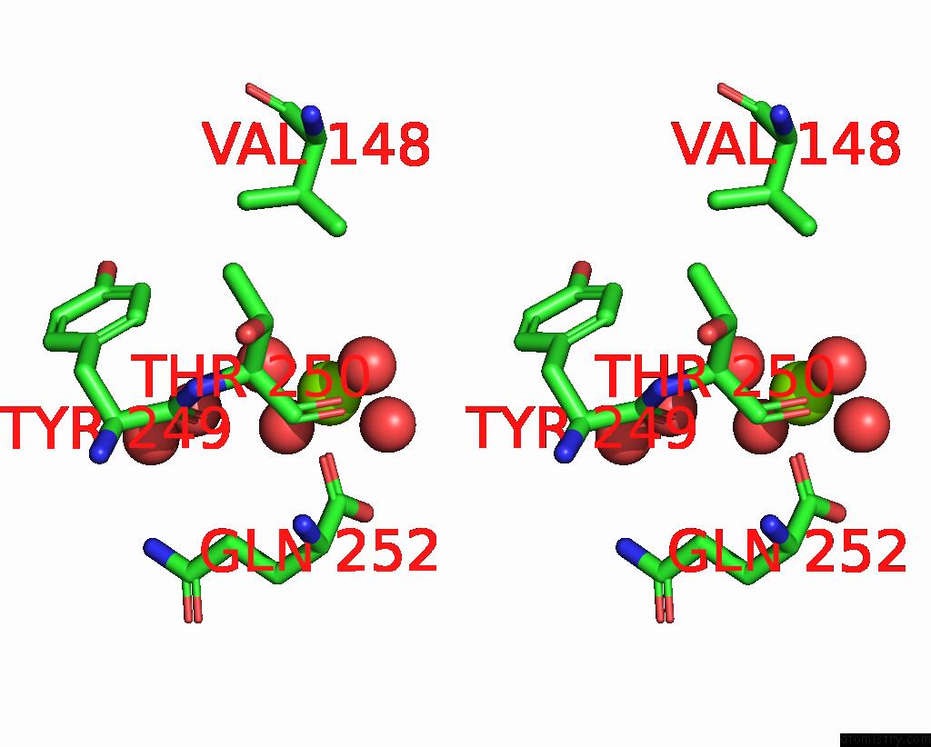
Stereo pair view
Mono view
Stereo pair view
A full contact list of Magnesium with other atoms in the Mg binding
site number 1 of Crystal Structure of Ketoreductase From Lactobacillus Kefir, E145S Mutant within 5.0Å range:
|
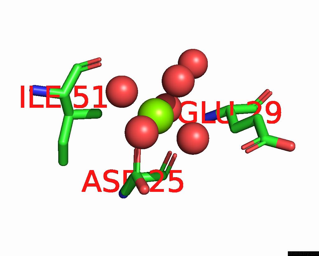
Magnesium binding site 2 out of 2 in 4rf5
Go back to
Magnesium binding site 2 out
of 2 in the Crystal Structure of Ketoreductase From Lactobacillus Kefir, E145S Mutant
Mono view
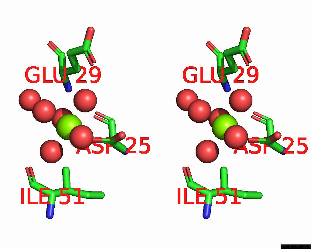
Stereo pair view
Mono view
Stereo pair view
A full contact list of Magnesium with other atoms in the Mg binding
site number 2 of Crystal Structure of Ketoreductase From Lactobacillus Kefir, E145S Mutant within 5.0Å range:
|
Reference:
E.L.Noey,
N.Tibrewal,
G.Jimenez-Oses,
S.Osuna,
J.Park,
C.M.Bond,
D.Cascio,
J.Liang,
X.Zhang,
G.W.Huisman,
Y.Tang,
K.N.Houk.
Origins of Stereoselectivity in Evolved Ketoreductases. Proc.Natl.Acad.Sci.Usa V. 112 E7065 2015.
ISSN: ISSN 0027-8424
PubMed: 26644568
DOI: 10.1073/PNAS.1507910112
Page generated: Tue Aug 20 03:04:15 2024
ISSN: ISSN 0027-8424
PubMed: 26644568
DOI: 10.1073/PNAS.1507910112
Last articles
Zn in 9J0NZn in 9J0O
Zn in 9J0P
Zn in 9FJX
Zn in 9EKB
Zn in 9C0F
Zn in 9CAH
Zn in 9CH0
Zn in 9CH3
Zn in 9CH1