Magnesium »
PDB 4rcz-4rkf »
4rgv »
Magnesium in PDB 4rgv: Crystal Structure of the Methanocaldococcus Jannaschii G1PDH
Enzymatic activity of Crystal Structure of the Methanocaldococcus Jannaschii G1PDH
All present enzymatic activity of Crystal Structure of the Methanocaldococcus Jannaschii G1PDH:
1.1.1.261;
1.1.1.261;
Protein crystallography data
The structure of Crystal Structure of the Methanocaldococcus Jannaschii G1PDH, PDB code: 4rgv
was solved by
V.Carbone,
R.S.Ronimus,
L.R.Schofield,
A.J.Sutherland-Smith,
with X-Ray Crystallography technique. A brief refinement statistics is given in the table below:
| Resolution Low / High (Å) | 46.07 / 2.45 |
| Space group | P 1 21 1 |
| Cell size a, b, c (Å), α, β, γ (°) | 49.765, 59.552, 119.825, 90.00, 90.39, 90.00 |
| R / Rfree (%) | 19.3 / 23.1 |
Other elements in 4rgv:
The structure of Crystal Structure of the Methanocaldococcus Jannaschii G1PDH also contains other interesting chemical elements:
| Zinc | (Zn) | 2 atoms |
Magnesium Binding Sites:
The binding sites of Magnesium atom in the Crystal Structure of the Methanocaldococcus Jannaschii G1PDH
(pdb code 4rgv). This binding sites where shown within
5.0 Angstroms radius around Magnesium atom.
In total 2 binding sites of Magnesium where determined in the Crystal Structure of the Methanocaldococcus Jannaschii G1PDH, PDB code: 4rgv:
Jump to Magnesium binding site number: 1; 2;
In total 2 binding sites of Magnesium where determined in the Crystal Structure of the Methanocaldococcus Jannaschii G1PDH, PDB code: 4rgv:
Jump to Magnesium binding site number: 1; 2;
Magnesium binding site 1 out of 2 in 4rgv
Go back to
Magnesium binding site 1 out
of 2 in the Crystal Structure of the Methanocaldococcus Jannaschii G1PDH
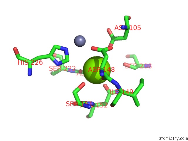
Mono view
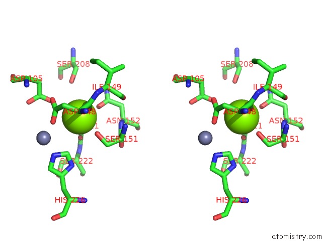
Stereo pair view
Mono view
Stereo pair view
A full contact list of Magnesium with other atoms in the Mg binding
site number 1 of Crystal Structure of the Methanocaldococcus Jannaschii G1PDH within 5.0Å range:
|
Magnesium binding site 2 out of 2 in 4rgv
Go back to
Magnesium binding site 2 out
of 2 in the Crystal Structure of the Methanocaldococcus Jannaschii G1PDH
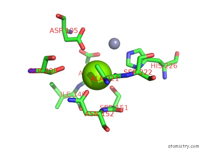
Mono view
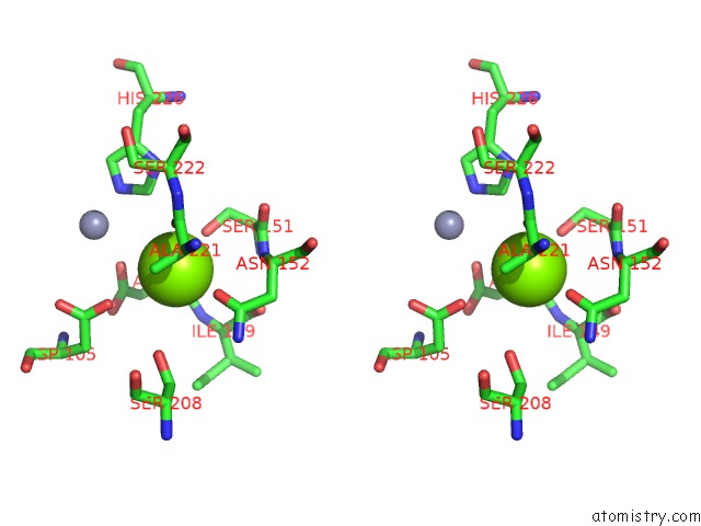
Stereo pair view
Mono view
Stereo pair view
A full contact list of Magnesium with other atoms in the Mg binding
site number 2 of Crystal Structure of the Methanocaldococcus Jannaschii G1PDH within 5.0Å range:
|
Reference:
V.Carbone,
L.R.Schofield,
Y.Zhang,
C.Sang,
D.Dey,
I.M.Hannus,
W.F.Martin,
A.J.Sutherland-Smith,
R.S.Ronimus.
Structure and Evolution of the Archaeal Lipid Synthesis Enzyme Sn-Glycerol-1-Phosphate Dehydrogenase. J.Biol.Chem. V. 290 21690 2015.
ISSN: ISSN 0021-9258
PubMed: 26175150
DOI: 10.1074/JBC.M115.647461
Page generated: Tue Aug 20 03:04:45 2024
ISSN: ISSN 0021-9258
PubMed: 26175150
DOI: 10.1074/JBC.M115.647461
Last articles
Zn in 9MJ5Zn in 9HNW
Zn in 9G0L
Zn in 9FNE
Zn in 9DZN
Zn in 9E0I
Zn in 9D32
Zn in 9DAK
Zn in 8ZXC
Zn in 8ZUF