Magnesium »
PDB 4rkq-4rri »
4rop »
Magnesium in PDB 4rop: Crystal Structure of Enolase From Synechococcus Elongatus
Enzymatic activity of Crystal Structure of Enolase From Synechococcus Elongatus
All present enzymatic activity of Crystal Structure of Enolase From Synechococcus Elongatus:
4.2.1.11;
4.2.1.11;
Protein crystallography data
The structure of Crystal Structure of Enolase From Synechococcus Elongatus, PDB code: 4rop
was solved by
J.M.Gonzalez,
R.Marti-Arbona,
C.J.Unkefer,
with X-Ray Crystallography technique. A brief refinement statistics is given in the table below:
| Resolution Low / High (Å) | 37.75 / 2.05 |
| Space group | I 4 2 2 |
| Cell size a, b, c (Å), α, β, γ (°) | 164.282, 164.282, 75.500, 90.00, 90.00, 90.00 |
| R / Rfree (%) | 19.9 / 22.8 |
Other elements in 4rop:
The structure of Crystal Structure of Enolase From Synechococcus Elongatus also contains other interesting chemical elements:
| Chlorine | (Cl) | 1 atom |
| Calcium | (Ca) | 4 atoms |
Magnesium Binding Sites:
The binding sites of Magnesium atom in the Crystal Structure of Enolase From Synechococcus Elongatus
(pdb code 4rop). This binding sites where shown within
5.0 Angstroms radius around Magnesium atom.
In total only one binding site of Magnesium was determined in the Crystal Structure of Enolase From Synechococcus Elongatus, PDB code: 4rop:
In total only one binding site of Magnesium was determined in the Crystal Structure of Enolase From Synechococcus Elongatus, PDB code: 4rop:
Magnesium binding site 1 out of 1 in 4rop
Go back to
Magnesium binding site 1 out
of 1 in the Crystal Structure of Enolase From Synechococcus Elongatus
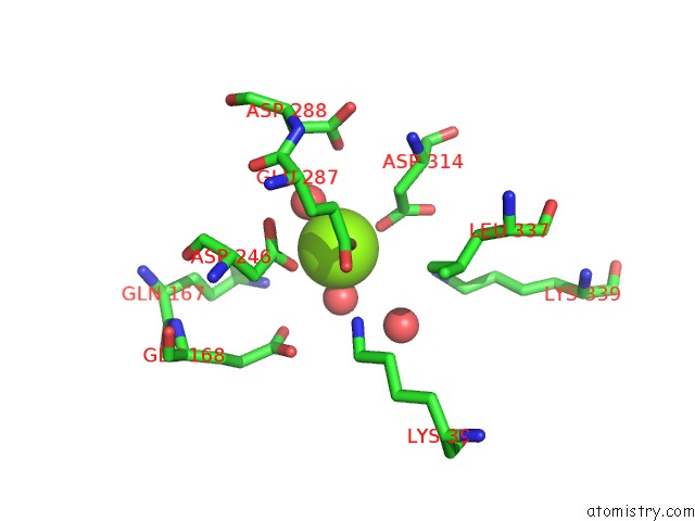
Mono view
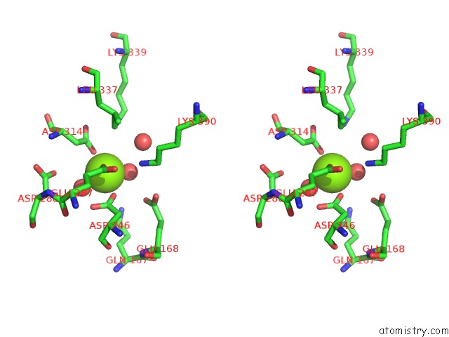
Stereo pair view
Mono view
Stereo pair view
A full contact list of Magnesium with other atoms in the Mg binding
site number 1 of Crystal Structure of Enolase From Synechococcus Elongatus within 5.0Å range:
|
Reference:
J.M.Gonzalez,
R.Marti-Arbona,
C.J.Unkefer.
Crystal Structure of Enolase From Synechococcus Elongatus To Be Published.
Page generated: Tue Aug 20 03:15:31 2024
Last articles
Cl in 7SFICl in 7SF3
Cl in 7SBJ
Cl in 7SEL
Cl in 7SET
Cl in 7SDR
Cl in 7SCI
Cl in 7SB6
Cl in 7SAK
Cl in 7SAC