Magnesium »
PDB 4tyw-4u9h »
4u0m »
Magnesium in PDB 4u0m: Structure of the Vibrio Cholerae Di-Nucleotide Cyclase (Dncv) Mutant D193N in Complex with Atp, Gtp and 5MTHFGLU2
Enzymatic activity of Structure of the Vibrio Cholerae Di-Nucleotide Cyclase (Dncv) Mutant D193N in Complex with Atp, Gtp and 5MTHFGLU2
All present enzymatic activity of Structure of the Vibrio Cholerae Di-Nucleotide Cyclase (Dncv) Mutant D193N in Complex with Atp, Gtp and 5MTHFGLU2:
2.7.7.86;
2.7.7.86;
Protein crystallography data
The structure of Structure of the Vibrio Cholerae Di-Nucleotide Cyclase (Dncv) Mutant D193N in Complex with Atp, Gtp and 5MTHFGLU2, PDB code: 4u0m
was solved by
D.Zhu,
Y.Xiang,
with X-Ray Crystallography technique. A brief refinement statistics is given in the table below:
| Resolution Low / High (Å) | 42.57 / 2.30 |
| Space group | P 1 21 1 |
| Cell size a, b, c (Å), α, β, γ (°) | 69.898, 59.860, 104.185, 90.00, 95.55, 90.00 |
| R / Rfree (%) | 19.9 / 25.3 |
Magnesium Binding Sites:
The binding sites of Magnesium atom in the Structure of the Vibrio Cholerae Di-Nucleotide Cyclase (Dncv) Mutant D193N in Complex with Atp, Gtp and 5MTHFGLU2
(pdb code 4u0m). This binding sites where shown within
5.0 Angstroms radius around Magnesium atom.
In total 2 binding sites of Magnesium where determined in the Structure of the Vibrio Cholerae Di-Nucleotide Cyclase (Dncv) Mutant D193N in Complex with Atp, Gtp and 5MTHFGLU2, PDB code: 4u0m:
Jump to Magnesium binding site number: 1; 2;
In total 2 binding sites of Magnesium where determined in the Structure of the Vibrio Cholerae Di-Nucleotide Cyclase (Dncv) Mutant D193N in Complex with Atp, Gtp and 5MTHFGLU2, PDB code: 4u0m:
Jump to Magnesium binding site number: 1; 2;
Magnesium binding site 1 out of 2 in 4u0m
Go back to
Magnesium binding site 1 out
of 2 in the Structure of the Vibrio Cholerae Di-Nucleotide Cyclase (Dncv) Mutant D193N in Complex with Atp, Gtp and 5MTHFGLU2

Mono view
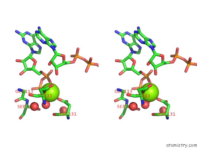
Stereo pair view
Mono view
Stereo pair view
A full contact list of Magnesium with other atoms in the Mg binding
site number 1 of Structure of the Vibrio Cholerae Di-Nucleotide Cyclase (Dncv) Mutant D193N in Complex with Atp, Gtp and 5MTHFGLU2 within 5.0Å range:
|
Magnesium binding site 2 out of 2 in 4u0m
Go back to
Magnesium binding site 2 out
of 2 in the Structure of the Vibrio Cholerae Di-Nucleotide Cyclase (Dncv) Mutant D193N in Complex with Atp, Gtp and 5MTHFGLU2

Mono view
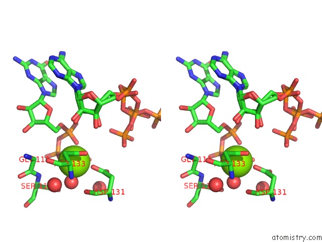
Stereo pair view
Mono view
Stereo pair view
A full contact list of Magnesium with other atoms in the Mg binding
site number 2 of Structure of the Vibrio Cholerae Di-Nucleotide Cyclase (Dncv) Mutant D193N in Complex with Atp, Gtp and 5MTHFGLU2 within 5.0Å range:
|
Reference:
D.Zhu,
L.Wang,
G.Shang,
X.Liu,
J.Zhu,
D.Lu,
L.Wang,
B.Kan,
J.R.Zhang,
Y.Xiang.
Structural Biochemistry of A Vibrio Cholerae Dinucleotide Cyclase Reveals Cyclase Activity Regulation By Folates. Mol.Cell V. 55 931 2014.
ISSN: ISSN 1097-2765
PubMed: 25201413
DOI: 10.1016/J.MOLCEL.2014.08.001
Page generated: Tue Aug 20 04:22:36 2024
ISSN: ISSN 1097-2765
PubMed: 25201413
DOI: 10.1016/J.MOLCEL.2014.08.001
Last articles
Fe in 2YXOFe in 2YRS
Fe in 2YXC
Fe in 2YNM
Fe in 2YVJ
Fe in 2YP1
Fe in 2YU2
Fe in 2YU1
Fe in 2YQB
Fe in 2YOO