Magnesium »
PDB 4tyq-4u98 »
4u5p »
Magnesium in PDB 4u5p: Crystal Structure of Native Rhcc (YP_702633.1) From Rhodococcus Jostii RHA1 at 1.78 Angstrom
Protein crystallography data
The structure of Crystal Structure of Native Rhcc (YP_702633.1) From Rhodococcus Jostii RHA1 at 1.78 Angstrom, PDB code: 4u5p
was solved by
H.Poddar,
H.J.Rozeboom,
A.M.W.H.Thunnissen,
with X-Ray Crystallography technique. A brief refinement statistics is given in the table below:
| Resolution Low / High (Å) | 46.35 / 1.78 |
| Space group | C 2 2 21 |
| Cell size a, b, c (Å), α, β, γ (°) | 54.690, 98.550, 188.530, 90.00, 90.00, 90.00 |
| R / Rfree (%) | 15 / 18.7 |
Magnesium Binding Sites:
The binding sites of Magnesium atom in the Crystal Structure of Native Rhcc (YP_702633.1) From Rhodococcus Jostii RHA1 at 1.78 Angstrom
(pdb code 4u5p). This binding sites where shown within
5.0 Angstroms radius around Magnesium atom.
In total only one binding site of Magnesium was determined in the Crystal Structure of Native Rhcc (YP_702633.1) From Rhodococcus Jostii RHA1 at 1.78 Angstrom, PDB code: 4u5p:
In total only one binding site of Magnesium was determined in the Crystal Structure of Native Rhcc (YP_702633.1) From Rhodococcus Jostii RHA1 at 1.78 Angstrom, PDB code: 4u5p:
Magnesium binding site 1 out of 1 in 4u5p
Go back to
Magnesium binding site 1 out
of 1 in the Crystal Structure of Native Rhcc (YP_702633.1) From Rhodococcus Jostii RHA1 at 1.78 Angstrom
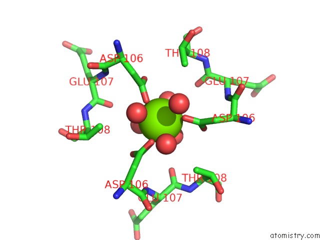
Mono view
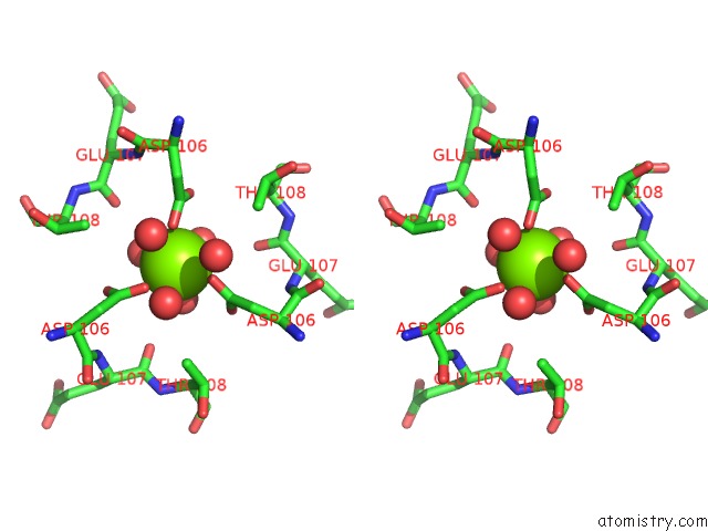
Stereo pair view
Mono view
Stereo pair view
A full contact list of Magnesium with other atoms in the Mg binding
site number 1 of Crystal Structure of Native Rhcc (YP_702633.1) From Rhodococcus Jostii RHA1 at 1.78 Angstrom within 5.0Å range:
|
Reference:
B.J.Baas,
H.Poddar,
E.M.Geertsema,
H.J.Rozeboom,
M.P.De Vries,
H.P.Permentier,
A.M.Thunnissen,
G.J.Poelarends.
Functional and Structural Characterization of An Unusual Cofactor-Independent Oxygenase. Biochemistry V. 54 1219 2015.
ISSN: ISSN 0006-2960
PubMed: 25565350
DOI: 10.1021/BI501200J
Page generated: Tue Aug 20 04:26:05 2024
ISSN: ISSN 0006-2960
PubMed: 25565350
DOI: 10.1021/BI501200J
Last articles
Zn in 9J0NZn in 9J0O
Zn in 9J0P
Zn in 9FJX
Zn in 9EKB
Zn in 9C0F
Zn in 9CAH
Zn in 9CH0
Zn in 9CH3
Zn in 9CH1