Magnesium »
PDB 4u9h-4ukd »
4uau »
Magnesium in PDB 4uau: Crystal Structure of Cbby (Mutant D10N) From Rhodobacter Sphaeroides in Complex with Xylulose-(1,5)Bisphosphate, Crystal Form II
Protein crystallography data
The structure of Crystal Structure of Cbby (Mutant D10N) From Rhodobacter Sphaeroides in Complex with Xylulose-(1,5)Bisphosphate, Crystal Form II, PDB code: 4uau
was solved by
A.Bracher,
A.Sharma,
A.Starling-Windhof,
F.U.Hartl,
M.Hayer-Hartl,
with X-Ray Crystallography technique. A brief refinement statistics is given in the table below:
| Resolution Low / High (Å) | 30.00 / 1.45 |
| Space group | P 21 21 21 |
| Cell size a, b, c (Å), α, β, γ (°) | 51.288, 69.767, 125.793, 90.00, 90.00, 90.00 |
| R / Rfree (%) | 17.9 / 21.1 |
Magnesium Binding Sites:
The binding sites of Magnesium atom in the Crystal Structure of Cbby (Mutant D10N) From Rhodobacter Sphaeroides in Complex with Xylulose-(1,5)Bisphosphate, Crystal Form II
(pdb code 4uau). This binding sites where shown within
5.0 Angstroms radius around Magnesium atom.
In total 2 binding sites of Magnesium where determined in the Crystal Structure of Cbby (Mutant D10N) From Rhodobacter Sphaeroides in Complex with Xylulose-(1,5)Bisphosphate, Crystal Form II, PDB code: 4uau:
Jump to Magnesium binding site number: 1; 2;
In total 2 binding sites of Magnesium where determined in the Crystal Structure of Cbby (Mutant D10N) From Rhodobacter Sphaeroides in Complex with Xylulose-(1,5)Bisphosphate, Crystal Form II, PDB code: 4uau:
Jump to Magnesium binding site number: 1; 2;
Magnesium binding site 1 out of 2 in 4uau
Go back to
Magnesium binding site 1 out
of 2 in the Crystal Structure of Cbby (Mutant D10N) From Rhodobacter Sphaeroides in Complex with Xylulose-(1,5)Bisphosphate, Crystal Form II

Mono view
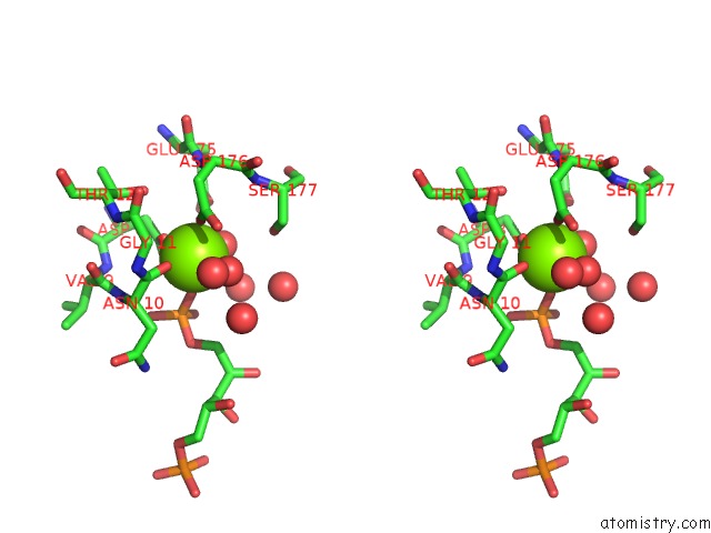
Stereo pair view
Mono view
Stereo pair view
A full contact list of Magnesium with other atoms in the Mg binding
site number 1 of Crystal Structure of Cbby (Mutant D10N) From Rhodobacter Sphaeroides in Complex with Xylulose-(1,5)Bisphosphate, Crystal Form II within 5.0Å range:
|
Magnesium binding site 2 out of 2 in 4uau
Go back to
Magnesium binding site 2 out
of 2 in the Crystal Structure of Cbby (Mutant D10N) From Rhodobacter Sphaeroides in Complex with Xylulose-(1,5)Bisphosphate, Crystal Form II
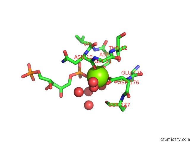
Mono view

Stereo pair view
Mono view
Stereo pair view
A full contact list of Magnesium with other atoms in the Mg binding
site number 2 of Crystal Structure of Cbby (Mutant D10N) From Rhodobacter Sphaeroides in Complex with Xylulose-(1,5)Bisphosphate, Crystal Form II within 5.0Å range:
|
Reference:
A.Bracher,
A.Sharma,
A.Starling-Windhof,
F.U.Hartl,
M.Hayer-Hartl.
Degradation of Potent Rubisco Inhibitor By Selective Sugar Phosphatase Nat.Plants 2015.
DOI: 10.1038/NPLANTS.2014.2
Page generated: Tue Aug 20 04:34:12 2024
DOI: 10.1038/NPLANTS.2014.2
Last articles
Zn in 9J0NZn in 9J0O
Zn in 9J0P
Zn in 9FJX
Zn in 9EKB
Zn in 9C0F
Zn in 9CAH
Zn in 9CH0
Zn in 9CH3
Zn in 9CH1