Magnesium »
PDB 4u9i-4um5 »
4uhk »
Magnesium in PDB 4uhk: Crystal Structure of the Receiver Domain of Cpxr From E. Coli (Phosphorylated)
Protein crystallography data
The structure of Crystal Structure of the Receiver Domain of Cpxr From E. Coli (Phosphorylated), PDB code: 4uhk
was solved by
A.E.Mechaly,
P.M.A.Alzari,
with X-Ray Crystallography technique. A brief refinement statistics is given in the table below:
| Resolution Low / High (Å) | 45.38 / 2.60 |
| Space group | P 21 21 2 |
| Cell size a, b, c (Å), α, β, γ (°) | 86.831, 74.144, 76.450, 90.00, 90.00, 90.00 |
| R / Rfree (%) | 18.6 / 23.3 |
Magnesium Binding Sites:
The binding sites of Magnesium atom in the Crystal Structure of the Receiver Domain of Cpxr From E. Coli (Phosphorylated)
(pdb code 4uhk). This binding sites where shown within
5.0 Angstroms radius around Magnesium atom.
In total 3 binding sites of Magnesium where determined in the Crystal Structure of the Receiver Domain of Cpxr From E. Coli (Phosphorylated), PDB code: 4uhk:
Jump to Magnesium binding site number: 1; 2; 3;
In total 3 binding sites of Magnesium where determined in the Crystal Structure of the Receiver Domain of Cpxr From E. Coli (Phosphorylated), PDB code: 4uhk:
Jump to Magnesium binding site number: 1; 2; 3;
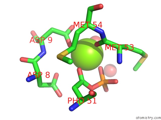
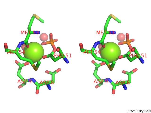
Magnesium binding site 1 out of 3 in 4uhk
Go back to
Magnesium binding site 1 out
of 3 in the Crystal Structure of the Receiver Domain of Cpxr From E. Coli (Phosphorylated)
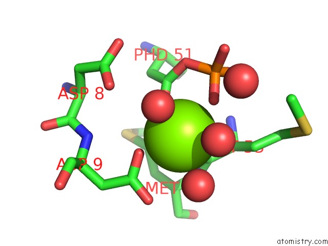
Mono view
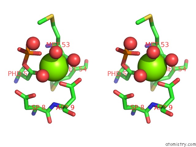
Stereo pair view
Mono view
Stereo pair view
A full contact list of Magnesium with other atoms in the Mg binding
site number 1 of Crystal Structure of the Receiver Domain of Cpxr From E. Coli (Phosphorylated) within 5.0Å range:
|
Magnesium binding site 2 out of 3 in 4uhk
Go back to
Magnesium binding site 2 out
of 3 in the Crystal Structure of the Receiver Domain of Cpxr From E. Coli (Phosphorylated)
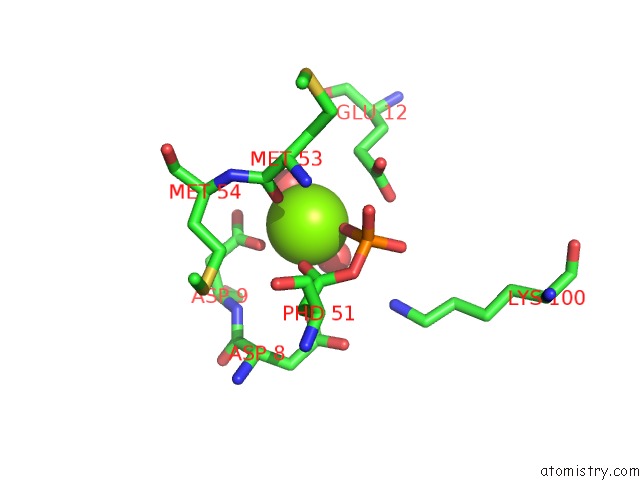
Mono view
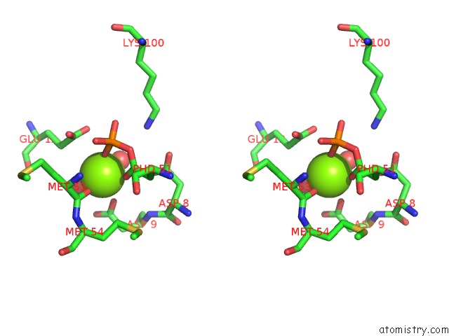
Stereo pair view
Mono view
Stereo pair view
A full contact list of Magnesium with other atoms in the Mg binding
site number 2 of Crystal Structure of the Receiver Domain of Cpxr From E. Coli (Phosphorylated) within 5.0Å range:
|
Magnesium binding site 3 out of 3 in 4uhk
Go back to
Magnesium binding site 3 out
of 3 in the Crystal Structure of the Receiver Domain of Cpxr From E. Coli (Phosphorylated)
Mono view
Stereo pair view
Mono view
Stereo pair view
A full contact list of Magnesium with other atoms in the Mg binding
site number 3 of Crystal Structure of the Receiver Domain of Cpxr From E. Coli (Phosphorylated) within 5.0Å range:
|
Reference:
A.E.Mechaly,
S.Soto Diaz,
N.Sassoon,
A.Buschiazzo,
J.M.Betton,
P.M.Alzari.
Structural Coupling Between Autokinase and Phosphotransferase Reactions in A Bacterial Histidine Kinase. Structure V. 25 939 2017.
ISSN: ISSN 1878-4186
PubMed: 28552574
DOI: 10.1016/J.STR.2017.04.011
Page generated: Tue Aug 20 04:43:42 2024
ISSN: ISSN 1878-4186
PubMed: 28552574
DOI: 10.1016/J.STR.2017.04.011
Last articles
F in 4KCSF in 4KCR
F in 4KCN
F in 4KCO
F in 4KBI
F in 4KBA
F in 4KBC
F in 4K9H
F in 4KAI
F in 4KB8