Magnesium »
PDB 4um5-4uuw »
4uof »
Magnesium in PDB 4uof: Crystallographic Structure of Nucleoside Diphosphate Kinase From Litopenaeus Vannamei Complexed with Dadp
Enzymatic activity of Crystallographic Structure of Nucleoside Diphosphate Kinase From Litopenaeus Vannamei Complexed with Dadp
All present enzymatic activity of Crystallographic Structure of Nucleoside Diphosphate Kinase From Litopenaeus Vannamei Complexed with Dadp:
2.7.4.6;
2.7.4.6;
Protein crystallography data
The structure of Crystallographic Structure of Nucleoside Diphosphate Kinase From Litopenaeus Vannamei Complexed with Dadp, PDB code: 4uof
was solved by
A.A.Lopez-Zavala,
V.Stojanoff,
E.Rudino-Pinera,
R.R.Sotelo-Mundo,
with X-Ray Crystallography technique. A brief refinement statistics is given in the table below:
| Resolution Low / High (Å) | 19.854 / 2.10 |
| Space group | C 2 2 21 |
| Cell size a, b, c (Å), α, β, γ (°) | 70.110, 134.380, 104.760, 90.00, 90.00, 90.00 |
| R / Rfree (%) | 18.95 / 24.93 |
Magnesium Binding Sites:
The binding sites of Magnesium atom in the Crystallographic Structure of Nucleoside Diphosphate Kinase From Litopenaeus Vannamei Complexed with Dadp
(pdb code 4uof). This binding sites where shown within
5.0 Angstroms radius around Magnesium atom.
In total 3 binding sites of Magnesium where determined in the Crystallographic Structure of Nucleoside Diphosphate Kinase From Litopenaeus Vannamei Complexed with Dadp, PDB code: 4uof:
Jump to Magnesium binding site number: 1; 2; 3;
In total 3 binding sites of Magnesium where determined in the Crystallographic Structure of Nucleoside Diphosphate Kinase From Litopenaeus Vannamei Complexed with Dadp, PDB code: 4uof:
Jump to Magnesium binding site number: 1; 2; 3;
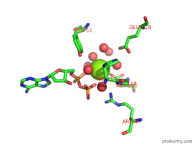
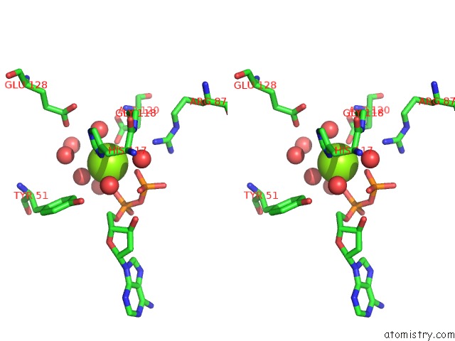
Magnesium binding site 1 out of 3 in 4uof
Go back to
Magnesium binding site 1 out
of 3 in the Crystallographic Structure of Nucleoside Diphosphate Kinase From Litopenaeus Vannamei Complexed with Dadp
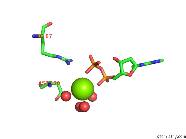
Mono view
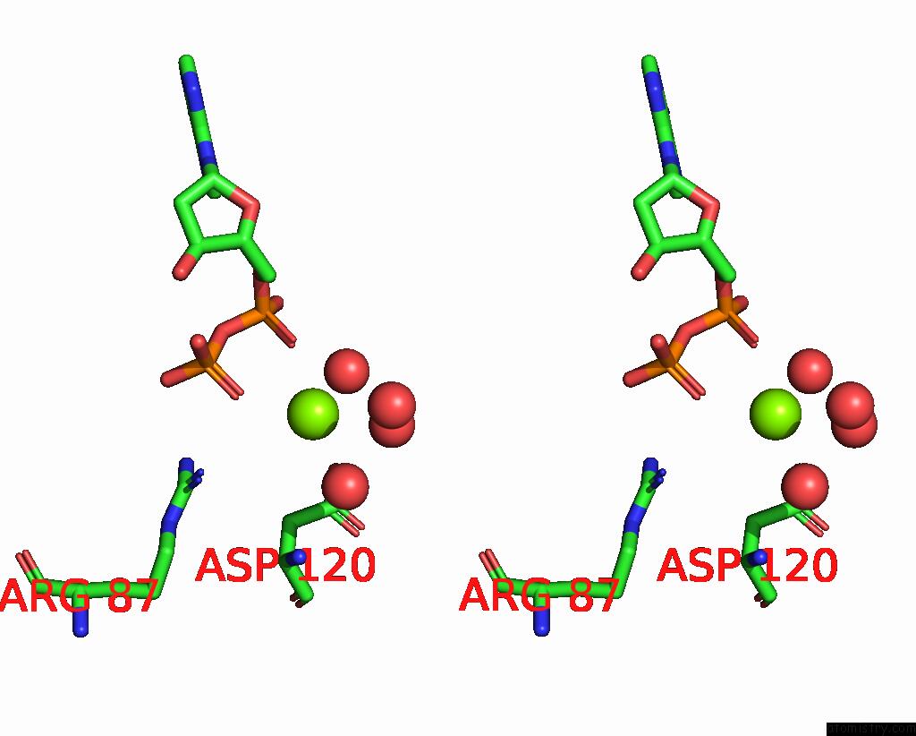
Stereo pair view
Mono view
Stereo pair view
A full contact list of Magnesium with other atoms in the Mg binding
site number 1 of Crystallographic Structure of Nucleoside Diphosphate Kinase From Litopenaeus Vannamei Complexed with Dadp within 5.0Å range:
|
Magnesium binding site 2 out of 3 in 4uof
Go back to
Magnesium binding site 2 out
of 3 in the Crystallographic Structure of Nucleoside Diphosphate Kinase From Litopenaeus Vannamei Complexed with Dadp
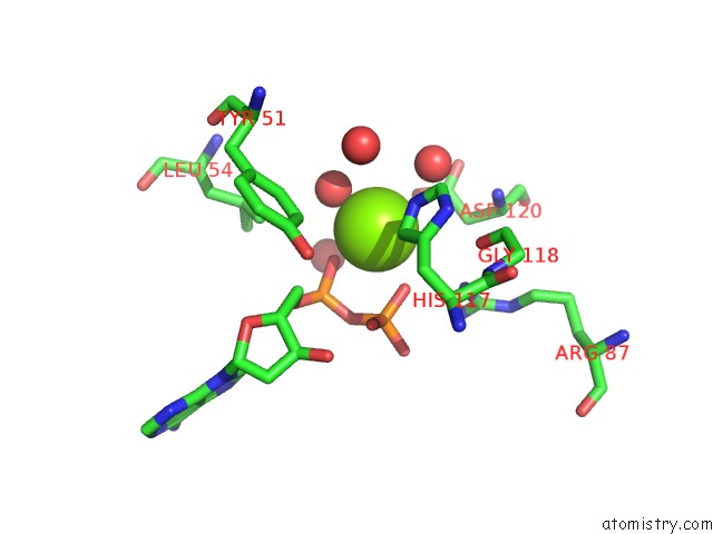
Mono view
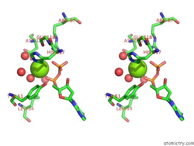
Stereo pair view
Mono view
Stereo pair view
A full contact list of Magnesium with other atoms in the Mg binding
site number 2 of Crystallographic Structure of Nucleoside Diphosphate Kinase From Litopenaeus Vannamei Complexed with Dadp within 5.0Å range:
|
Magnesium binding site 3 out of 3 in 4uof
Go back to
Magnesium binding site 3 out
of 3 in the Crystallographic Structure of Nucleoside Diphosphate Kinase From Litopenaeus Vannamei Complexed with Dadp
Mono view
Stereo pair view
Mono view
Stereo pair view
A full contact list of Magnesium with other atoms in the Mg binding
site number 3 of Crystallographic Structure of Nucleoside Diphosphate Kinase From Litopenaeus Vannamei Complexed with Dadp within 5.0Å range:
|
Reference:
A.A.Lopez-Zavala,
I.E.Quintero-Reyez,
J.S.Carrasco-Miranda,
V.Stojanoff,
A.Weichsel,
E.Rudino-Pinera,
R.R.Sotelo-Mundo.
Structure of Nucleoside Diphosphate Kinase From the Pacific Shrimp (Litopenaeus Vannamei) in Binary Complexes with Purine and Pyrimidine Nucleoside Diphosphates Acta Crystallogr.,Sect.F V. 79 1150 2014.
ISSN: ISSN 1744-3091
DOI: 10.1107/S2053230X1401557X
Page generated: Tue Aug 20 04:54:21 2024
ISSN: ISSN 1744-3091
DOI: 10.1107/S2053230X1401557X
Last articles
Zn in 9J0NZn in 9J0O
Zn in 9J0P
Zn in 9FJX
Zn in 9EKB
Zn in 9C0F
Zn in 9CAH
Zn in 9CH0
Zn in 9CH3
Zn in 9CH1