Magnesium »
PDB 4um7-4uux »
4uor »
Magnesium in PDB 4uor: Structure of Lipoteichoic Acid Synthase Ltas From Listeria Monocytogenes in Complex with Glycerol Phosphate
Protein crystallography data
The structure of Structure of Lipoteichoic Acid Synthase Ltas From Listeria Monocytogenes in Complex with Glycerol Phosphate, PDB code: 4uor
was solved by
I.Campeotto,
P.Freemont,
A.Grundling,
with X-Ray Crystallography technique. A brief refinement statistics is given in the table below:
| Resolution Low / High (Å) | 48.710 / 2.19 |
| Space group | P 21 21 21 |
| Cell size a, b, c (Å), α, β, γ (°) | 119.246, 119.625, 472.658, 90.00, 90.00, 90.00 |
| R / Rfree (%) | 17.82 / 21.36 |
Magnesium Binding Sites:
Pages:
>>> Page 1 <<< Page 2, Binding sites: 11 - 11;Binding sites:
The binding sites of Magnesium atom in the Structure of Lipoteichoic Acid Synthase Ltas From Listeria Monocytogenes in Complex with Glycerol Phosphate (pdb code 4uor). This binding sites where shown within 5.0 Angstroms radius around Magnesium atom.In total 11 binding sites of Magnesium where determined in the Structure of Lipoteichoic Acid Synthase Ltas From Listeria Monocytogenes in Complex with Glycerol Phosphate, PDB code: 4uor:
Jump to Magnesium binding site number: 1; 2; 3; 4; 5; 6; 7; 8; 9; 10;
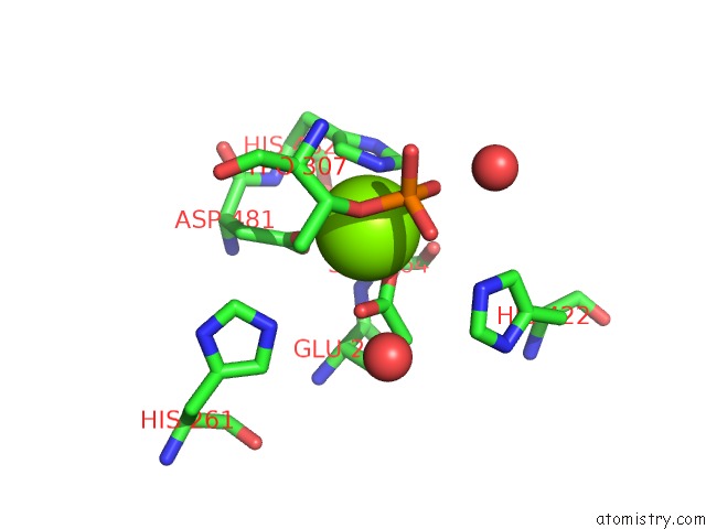
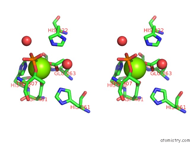
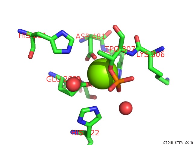
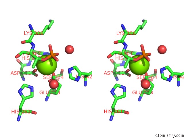
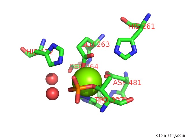
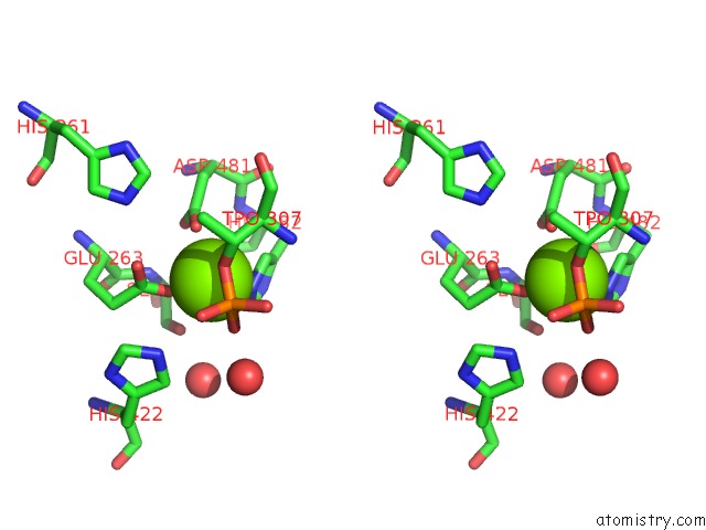
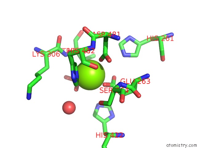
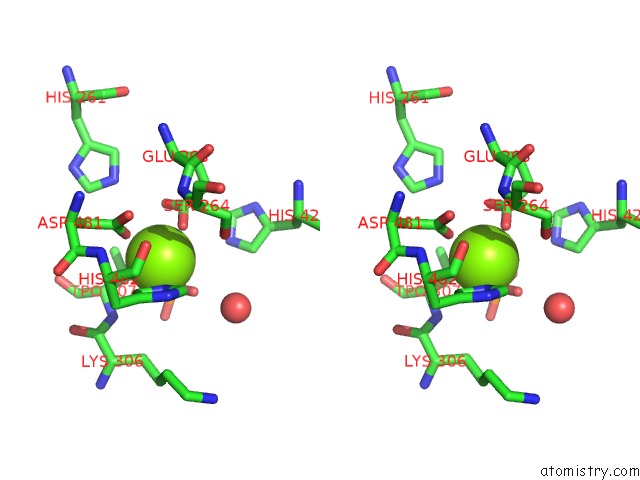
Magnesium binding site 1 out of 11 in 4uor
Go back to
Magnesium binding site 1 out
of 11 in the Structure of Lipoteichoic Acid Synthase Ltas From Listeria Monocytogenes in Complex with Glycerol Phosphate
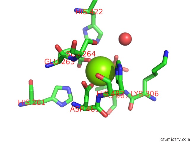
Mono view
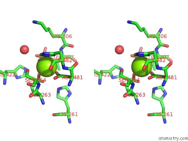
Stereo pair view
Mono view
Stereo pair view
A full contact list of Magnesium with other atoms in the Mg binding
site number 1 of Structure of Lipoteichoic Acid Synthase Ltas From Listeria Monocytogenes in Complex with Glycerol Phosphate within 5.0Å range:
|
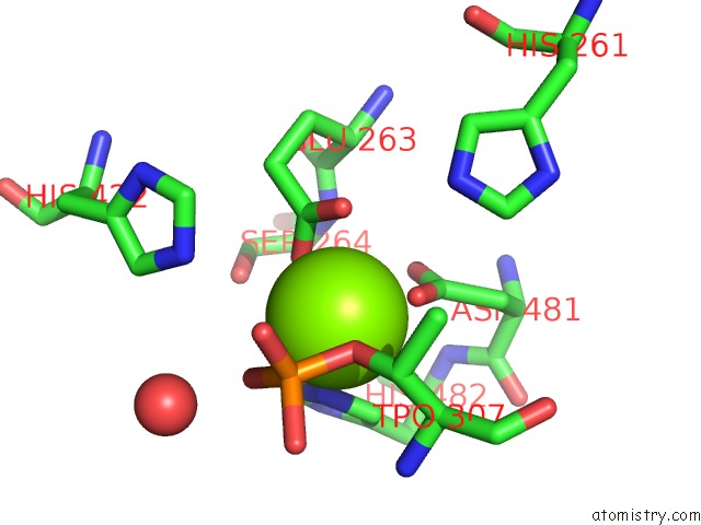
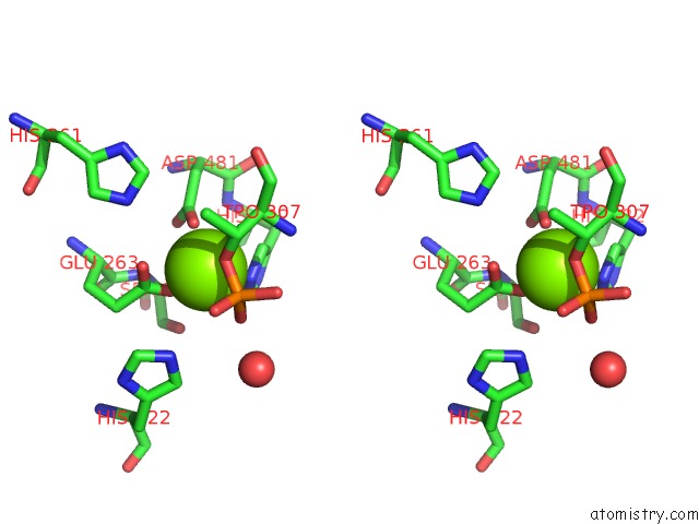
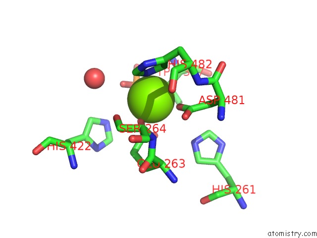
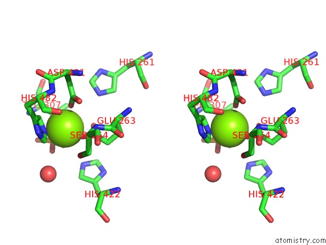
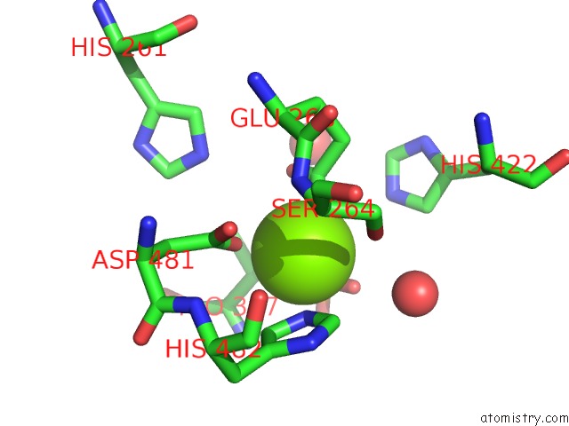
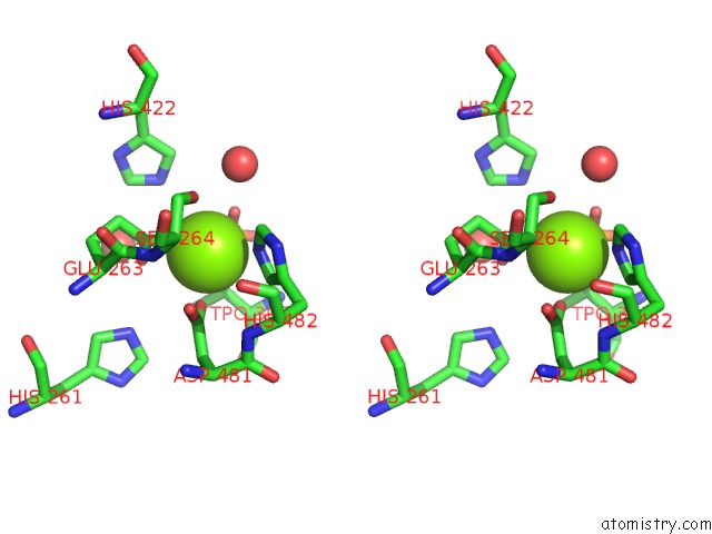
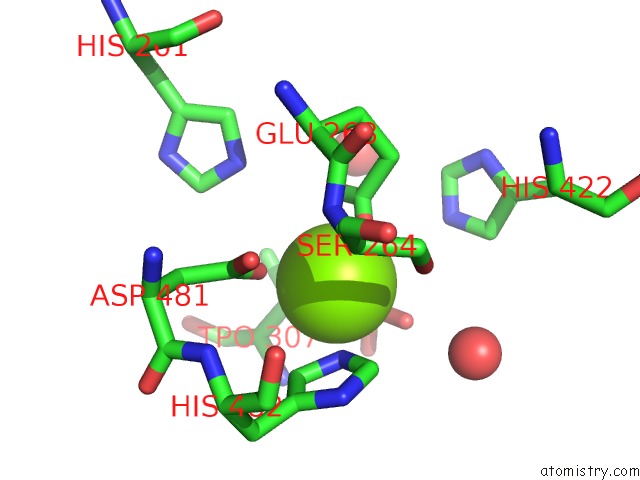
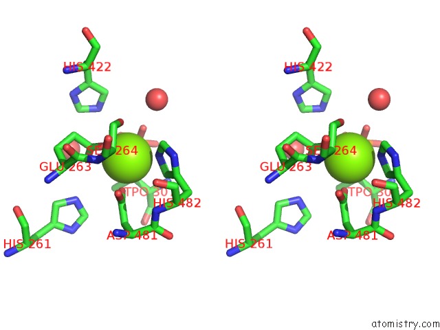
Magnesium binding site 2 out of 11 in 4uor
Go back to
Magnesium binding site 2 out
of 11 in the Structure of Lipoteichoic Acid Synthase Ltas From Listeria Monocytogenes in Complex with Glycerol Phosphate
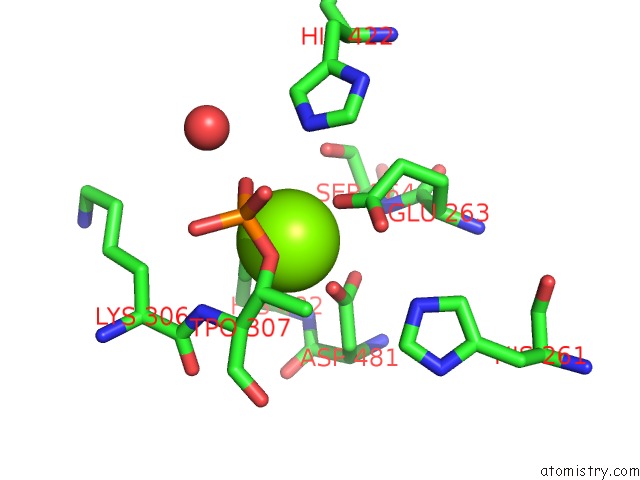
Mono view
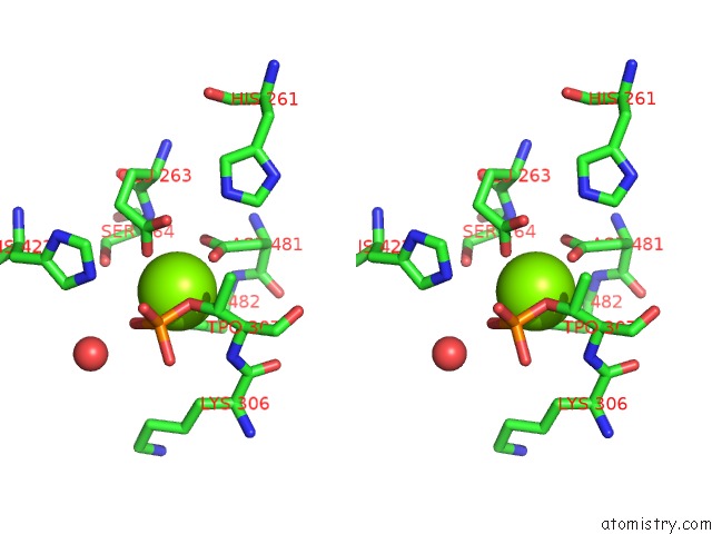
Stereo pair view
Mono view
Stereo pair view
A full contact list of Magnesium with other atoms in the Mg binding
site number 2 of Structure of Lipoteichoic Acid Synthase Ltas From Listeria Monocytogenes in Complex with Glycerol Phosphate within 5.0Å range:
|
Magnesium binding site 3 out of 11 in 4uor
Go back to
Magnesium binding site 3 out
of 11 in the Structure of Lipoteichoic Acid Synthase Ltas From Listeria Monocytogenes in Complex with Glycerol Phosphate
Mono view
Stereo pair view
Mono view
Stereo pair view
A full contact list of Magnesium with other atoms in the Mg binding
site number 3 of Structure of Lipoteichoic Acid Synthase Ltas From Listeria Monocytogenes in Complex with Glycerol Phosphate within 5.0Å range:
|
Magnesium binding site 4 out of 11 in 4uor
Go back to
Magnesium binding site 4 out
of 11 in the Structure of Lipoteichoic Acid Synthase Ltas From Listeria Monocytogenes in Complex with Glycerol Phosphate
Mono view
Stereo pair view
Mono view
Stereo pair view
A full contact list of Magnesium with other atoms in the Mg binding
site number 4 of Structure of Lipoteichoic Acid Synthase Ltas From Listeria Monocytogenes in Complex with Glycerol Phosphate within 5.0Å range:
|
Magnesium binding site 5 out of 11 in 4uor
Go back to
Magnesium binding site 5 out
of 11 in the Structure of Lipoteichoic Acid Synthase Ltas From Listeria Monocytogenes in Complex with Glycerol Phosphate
Mono view
Stereo pair view
Mono view
Stereo pair view
A full contact list of Magnesium with other atoms in the Mg binding
site number 5 of Structure of Lipoteichoic Acid Synthase Ltas From Listeria Monocytogenes in Complex with Glycerol Phosphate within 5.0Å range:
|
Magnesium binding site 6 out of 11 in 4uor
Go back to
Magnesium binding site 6 out
of 11 in the Structure of Lipoteichoic Acid Synthase Ltas From Listeria Monocytogenes in Complex with Glycerol Phosphate
Mono view
Stereo pair view
Mono view
Stereo pair view
A full contact list of Magnesium with other atoms in the Mg binding
site number 6 of Structure of Lipoteichoic Acid Synthase Ltas From Listeria Monocytogenes in Complex with Glycerol Phosphate within 5.0Å range:
|
Magnesium binding site 7 out of 11 in 4uor
Go back to
Magnesium binding site 7 out
of 11 in the Structure of Lipoteichoic Acid Synthase Ltas From Listeria Monocytogenes in Complex with Glycerol Phosphate
Mono view
Stereo pair view
Mono view
Stereo pair view
A full contact list of Magnesium with other atoms in the Mg binding
site number 7 of Structure of Lipoteichoic Acid Synthase Ltas From Listeria Monocytogenes in Complex with Glycerol Phosphate within 5.0Å range:
|
Magnesium binding site 8 out of 11 in 4uor
Go back to
Magnesium binding site 8 out
of 11 in the Structure of Lipoteichoic Acid Synthase Ltas From Listeria Monocytogenes in Complex with Glycerol Phosphate
Mono view
Stereo pair view
Mono view
Stereo pair view
A full contact list of Magnesium with other atoms in the Mg binding
site number 8 of Structure of Lipoteichoic Acid Synthase Ltas From Listeria Monocytogenes in Complex with Glycerol Phosphate within 5.0Å range:
|
Magnesium binding site 9 out of 11 in 4uor
Go back to
Magnesium binding site 9 out
of 11 in the Structure of Lipoteichoic Acid Synthase Ltas From Listeria Monocytogenes in Complex with Glycerol Phosphate
Mono view
Stereo pair view
Mono view
Stereo pair view
A full contact list of Magnesium with other atoms in the Mg binding
site number 9 of Structure of Lipoteichoic Acid Synthase Ltas From Listeria Monocytogenes in Complex with Glycerol Phosphate within 5.0Å range:
|
Magnesium binding site 10 out of 11 in 4uor
Go back to
Magnesium binding site 10 out
of 11 in the Structure of Lipoteichoic Acid Synthase Ltas From Listeria Monocytogenes in Complex with Glycerol Phosphate
Mono view
Stereo pair view
Mono view
Stereo pair view
A full contact list of Magnesium with other atoms in the Mg binding
site number 10 of Structure of Lipoteichoic Acid Synthase Ltas From Listeria Monocytogenes in Complex with Glycerol Phosphate within 5.0Å range:
|
Reference:
I.Campeotto,
M.G.Percy,
J.T.Macdonald,
A.Forster,
P.S.Freemont,
A.Grundling.
Structural and Mechanistic Insight Into the Listeria Monocytogenes Two-Enzyme Lipoteichoic Acid Synthesis System J.Biol.Chem. V. 289 28054 2014.
ISSN: ISSN 0021-9258
PubMed: 25128528
DOI: 10.1074/JBC.M114.590570
Page generated: Tue Aug 20 04:55:08 2024
ISSN: ISSN 0021-9258
PubMed: 25128528
DOI: 10.1074/JBC.M114.590570
Last articles
Zn in 9MJ5Zn in 9HNW
Zn in 9G0L
Zn in 9FNE
Zn in 9DZN
Zn in 9E0I
Zn in 9D32
Zn in 9DAK
Zn in 8ZXC
Zn in 8ZUF