Magnesium »
PDB 4w5q-4wfn »
4wfn »
Magnesium in PDB 4wfn: Crystal Structure of the Large Ribosomal Subunit (50S) of Deinococcus Radiodurans Containing A Three Residue Insertion in L22 in Complex with Erythromycin
Protein crystallography data
The structure of Crystal Structure of the Large Ribosomal Subunit (50S) of Deinococcus Radiodurans Containing A Three Residue Insertion in L22 in Complex with Erythromycin, PDB code: 4wfn
was solved by
I.Wekselman,
E.Zimmerman,
H.Rozenberg,
A.Bashan,
A.Yonath,
with X-Ray Crystallography technique. A brief refinement statistics is given in the table below:
| Resolution Low / High (Å) | 19.99 / 3.54 |
| Space group | I 2 2 2 |
| Cell size a, b, c (Å), α, β, γ (°) | 170.092, 411.588, 695.883, 90.00, 90.00, 90.00 |
| R / Rfree (%) | 23.4 / 28.2 |
Magnesium Binding Sites:
Pages:
>>> Page 1 <<< Page 2, Binding sites: 11 - 20; Page 3, Binding sites: 21 - 30; Page 4, Binding sites: 31 - 40; Page 5, Binding sites: 41 - 50; Page 6, Binding sites: 51 - 60; Page 7, Binding sites: 61 - 70;Binding sites:
The binding sites of Magnesium atom in the Crystal Structure of the Large Ribosomal Subunit (50S) of Deinococcus Radiodurans Containing A Three Residue Insertion in L22 in Complex with Erythromycin (pdb code 4wfn). This binding sites where shown within 5.0 Angstroms radius around Magnesium atom.In total 70 binding sites of Magnesium where determined in the Crystal Structure of the Large Ribosomal Subunit (50S) of Deinococcus Radiodurans Containing A Three Residue Insertion in L22 in Complex with Erythromycin, PDB code: 4wfn:
Jump to Magnesium binding site number: 1; 2; 3; 4; 5; 6; 7; 8; 9; 10;
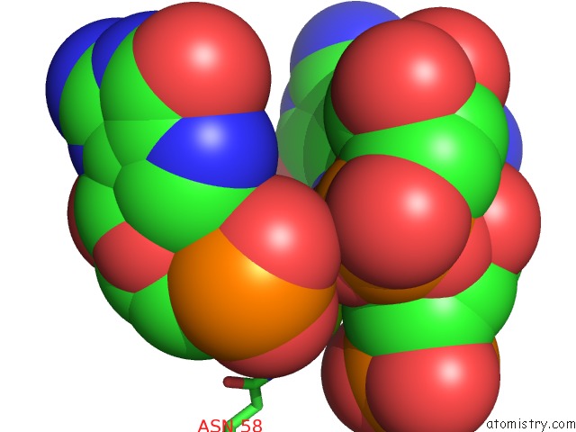
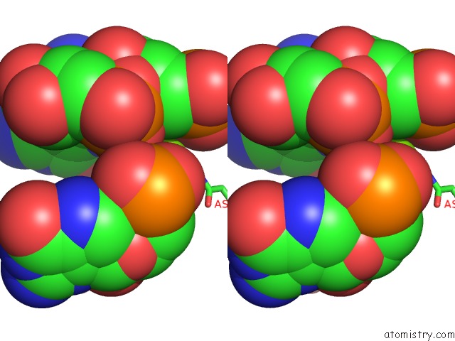
Magnesium binding site 1 out of 70 in 4wfn
Go back to
Magnesium binding site 1 out
of 70 in the Crystal Structure of the Large Ribosomal Subunit (50S) of Deinococcus Radiodurans Containing A Three Residue Insertion in L22 in Complex with Erythromycin
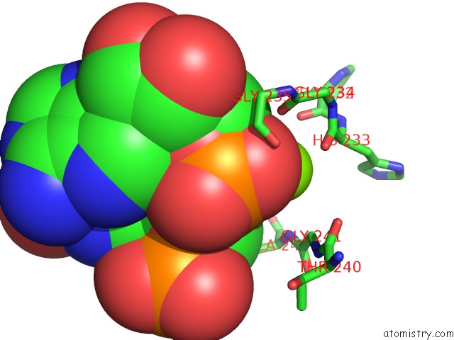
Mono view
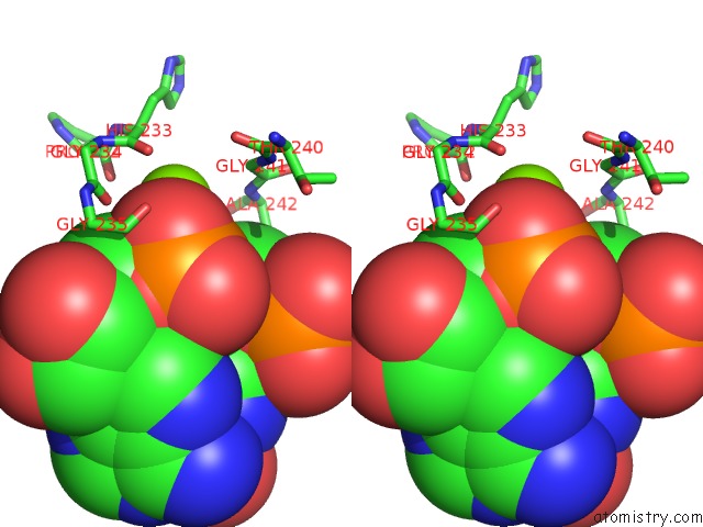
Stereo pair view
Mono view
Stereo pair view
A full contact list of Magnesium with other atoms in the Mg binding
site number 1 of Crystal Structure of the Large Ribosomal Subunit (50S) of Deinococcus Radiodurans Containing A Three Residue Insertion in L22 in Complex with Erythromycin within 5.0Å range:
|
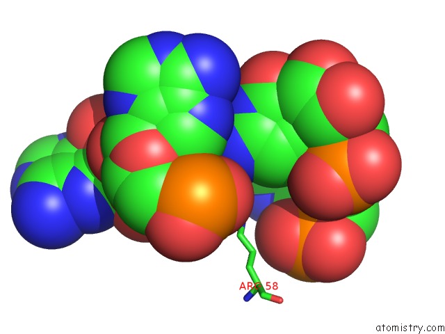
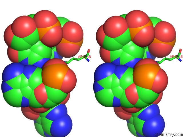
Magnesium binding site 2 out of 70 in 4wfn
Go back to
Magnesium binding site 2 out
of 70 in the Crystal Structure of the Large Ribosomal Subunit (50S) of Deinococcus Radiodurans Containing A Three Residue Insertion in L22 in Complex with Erythromycin
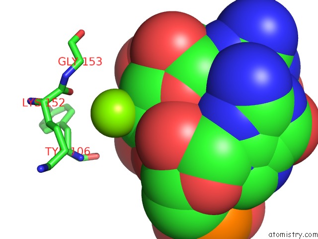
Mono view
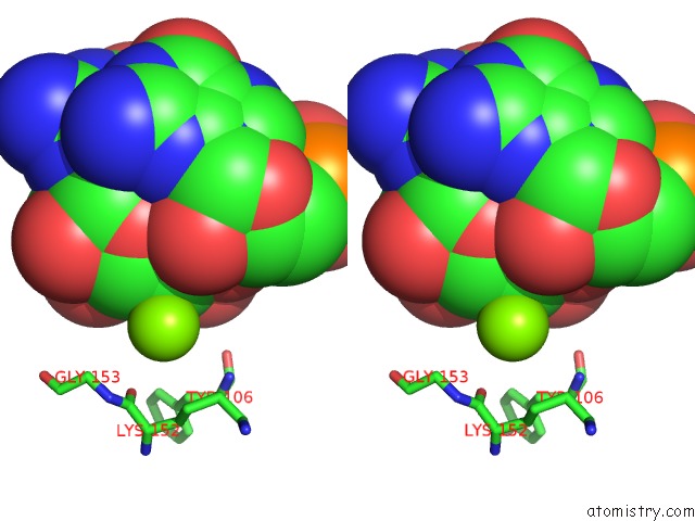
Stereo pair view
Mono view
Stereo pair view
A full contact list of Magnesium with other atoms in the Mg binding
site number 2 of Crystal Structure of the Large Ribosomal Subunit (50S) of Deinococcus Radiodurans Containing A Three Residue Insertion in L22 in Complex with Erythromycin within 5.0Å range:
|
Magnesium binding site 3 out of 70 in 4wfn
Go back to
Magnesium binding site 3 out
of 70 in the Crystal Structure of the Large Ribosomal Subunit (50S) of Deinococcus Radiodurans Containing A Three Residue Insertion in L22 in Complex with Erythromycin
Mono view
Stereo pair view
Mono view
Stereo pair view
A full contact list of Magnesium with other atoms in the Mg binding
site number 3 of Crystal Structure of the Large Ribosomal Subunit (50S) of Deinococcus Radiodurans Containing A Three Residue Insertion in L22 in Complex with Erythromycin within 5.0Å range:
|
Magnesium binding site 4 out of 70 in 4wfn
Go back to
Magnesium binding site 4 out
of 70 in the Crystal Structure of the Large Ribosomal Subunit (50S) of Deinococcus Radiodurans Containing A Three Residue Insertion in L22 in Complex with Erythromycin
Mono view
Stereo pair view
Mono view
Stereo pair view
A full contact list of Magnesium with other atoms in the Mg binding
site number 4 of Crystal Structure of the Large Ribosomal Subunit (50S) of Deinococcus Radiodurans Containing A Three Residue Insertion in L22 in Complex with Erythromycin within 5.0Å range:
|
Magnesium binding site 5 out of 70 in 4wfn
Go back to
Magnesium binding site 5 out
of 70 in the Crystal Structure of the Large Ribosomal Subunit (50S) of Deinococcus Radiodurans Containing A Three Residue Insertion in L22 in Complex with Erythromycin
Mono view

Stereo pair view
Mono view
Stereo pair view
A full contact list of Magnesium with other atoms in the Mg binding
site number 5 of Crystal Structure of the Large Ribosomal Subunit (50S) of Deinococcus Radiodurans Containing A Three Residue Insertion in L22 in Complex with Erythromycin within 5.0Å range:
|
Magnesium binding site 6 out of 70 in 4wfn
Go back to
Magnesium binding site 6 out
of 70 in the Crystal Structure of the Large Ribosomal Subunit (50S) of Deinococcus Radiodurans Containing A Three Residue Insertion in L22 in Complex with Erythromycin

Mono view
Stereo pair view
Mono view
Stereo pair view
A full contact list of Magnesium with other atoms in the Mg binding
site number 6 of Crystal Structure of the Large Ribosomal Subunit (50S) of Deinococcus Radiodurans Containing A Three Residue Insertion in L22 in Complex with Erythromycin within 5.0Å range:
|
Magnesium binding site 7 out of 70 in 4wfn
Go back to
Magnesium binding site 7 out
of 70 in the Crystal Structure of the Large Ribosomal Subunit (50S) of Deinococcus Radiodurans Containing A Three Residue Insertion in L22 in Complex with Erythromycin
Mono view
Stereo pair view
Mono view
Stereo pair view
A full contact list of Magnesium with other atoms in the Mg binding
site number 7 of Crystal Structure of the Large Ribosomal Subunit (50S) of Deinococcus Radiodurans Containing A Three Residue Insertion in L22 in Complex with Erythromycin within 5.0Å range:
|
Magnesium binding site 8 out of 70 in 4wfn
Go back to
Magnesium binding site 8 out
of 70 in the Crystal Structure of the Large Ribosomal Subunit (50S) of Deinococcus Radiodurans Containing A Three Residue Insertion in L22 in Complex with Erythromycin
Mono view
Stereo pair view
Mono view
Stereo pair view
A full contact list of Magnesium with other atoms in the Mg binding
site number 8 of Crystal Structure of the Large Ribosomal Subunit (50S) of Deinococcus Radiodurans Containing A Three Residue Insertion in L22 in Complex with Erythromycin within 5.0Å range:
|
Magnesium binding site 9 out of 70 in 4wfn
Go back to
Magnesium binding site 9 out
of 70 in the Crystal Structure of the Large Ribosomal Subunit (50S) of Deinococcus Radiodurans Containing A Three Residue Insertion in L22 in Complex with Erythromycin
Mono view
Stereo pair view
Mono view
Stereo pair view
A full contact list of Magnesium with other atoms in the Mg binding
site number 9 of Crystal Structure of the Large Ribosomal Subunit (50S) of Deinococcus Radiodurans Containing A Three Residue Insertion in L22 in Complex with Erythromycin within 5.0Å range:
|
Magnesium binding site 10 out of 70 in 4wfn
Go back to
Magnesium binding site 10 out
of 70 in the Crystal Structure of the Large Ribosomal Subunit (50S) of Deinococcus Radiodurans Containing A Three Residue Insertion in L22 in Complex with Erythromycin
Mono view
Stereo pair view
Mono view
Stereo pair view
A full contact list of Magnesium with other atoms in the Mg binding
site number 10 of Crystal Structure of the Large Ribosomal Subunit (50S) of Deinococcus Radiodurans Containing A Three Residue Insertion in L22 in Complex with Erythromycin within 5.0Å range:
|
Reference:
I.Wekselman,
E.Zimmerman,
C.Davidovich,
M.Belousoff,
D.Matzov,
M.Krupkin,
H.Rozenberg,
A.Bashan,
G.Friedlander,
J.Kjeldgaard,
H.Ingmer,
L.Lindahl,
J.M.Zengel,
A.Yonath.
The Ribosomal Protein UL22 Modulates the Shape of the Protein Exit Tunnel. Structure V. 25 1233 2017.
ISSN: ISSN 1878-4186
PubMed: 28689968
DOI: 10.1016/J.STR.2017.06.004
Page generated: Tue Aug 12 01:32:11 2025
ISSN: ISSN 1878-4186
PubMed: 28689968
DOI: 10.1016/J.STR.2017.06.004
Last articles
Mg in 5M50Mg in 5M3U
Mg in 5M4K
Mg in 5M41
Mg in 5M1T
Mg in 5M2T
Mg in 5M3F
Mg in 5LZQ
Mg in 5M2A
Mg in 5M1R