Magnesium »
PDB 4wfn-4wxe »
4wkj »
Magnesium in PDB 4wkj: Crystallographic Structure of A Dodecameric Rna-Dna Hybrid
Protein crystallography data
The structure of Crystallographic Structure of A Dodecameric Rna-Dna Hybrid, PDB code: 4wkj
was solved by
R.R.Davis,
N.M.Shaban,
F.W.Perrino,
T.Hollis,
with X-Ray Crystallography technique. A brief refinement statistics is given in the table below:
| Resolution Low / High (Å) | 26.27 / 2.80 |
| Space group | P 1 21 1 |
| Cell size a, b, c (Å), α, β, γ (°) | 46.649, 44.459, 83.346, 90.00, 105.27, 90.00 |
| R / Rfree (%) | 23.8 / 24.2 |
Magnesium Binding Sites:
The binding sites of Magnesium atom in the Crystallographic Structure of A Dodecameric Rna-Dna Hybrid
(pdb code 4wkj). This binding sites where shown within
5.0 Angstroms radius around Magnesium atom.
In total only one binding site of Magnesium was determined in the Crystallographic Structure of A Dodecameric Rna-Dna Hybrid, PDB code: 4wkj:
In total only one binding site of Magnesium was determined in the Crystallographic Structure of A Dodecameric Rna-Dna Hybrid, PDB code: 4wkj:
Magnesium binding site 1 out of 1 in 4wkj
Go back to
Magnesium binding site 1 out
of 1 in the Crystallographic Structure of A Dodecameric Rna-Dna Hybrid
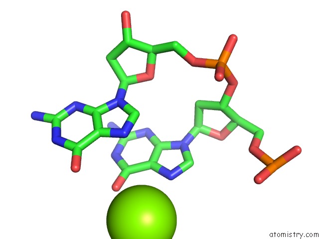
Mono view
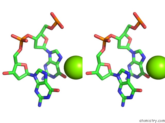
Stereo pair view
Mono view
Stereo pair view
A full contact list of Magnesium with other atoms in the Mg binding
site number 1 of Crystallographic Structure of A Dodecameric Rna-Dna Hybrid within 5.0Å range:
|
Reference:
R.R.Davis,
N.M.Shaban,
F.W.Perrino,
T.Hollis.
Crystal Structure of Rna-Dna Duplex Provides Insight Into Conformational Changes Induced By Rnase H Binding To Be Published.
Page generated: Tue Aug 20 13:52:47 2024
Last articles
Zn in 9J0NZn in 9J0O
Zn in 9J0P
Zn in 9FJX
Zn in 9EKB
Zn in 9C0F
Zn in 9CAH
Zn in 9CH0
Zn in 9CH3
Zn in 9CH1