Magnesium »
PDB 4wgi-4wxw »
4wud »
Magnesium in PDB 4wud: N-Terminal 43 kDa Fragment of the E. Coli Dna Gyrase B Subunit Grown From No Salt Condition
Enzymatic activity of N-Terminal 43 kDa Fragment of the E. Coli Dna Gyrase B Subunit Grown From No Salt Condition
All present enzymatic activity of N-Terminal 43 kDa Fragment of the E. Coli Dna Gyrase B Subunit Grown From No Salt Condition:
5.99.1.3;
5.99.1.3;
Protein crystallography data
The structure of N-Terminal 43 kDa Fragment of the E. Coli Dna Gyrase B Subunit Grown From No Salt Condition, PDB code: 4wud
was solved by
S.J.Hearnshaw,
T.T.Chung,
C.E.M.Stevenson,
A.Maxwell,
D.M.Lawson,
with X-Ray Crystallography technique. A brief refinement statistics is given in the table below:
| Resolution Low / High (Å) | 27.54 / 1.95 |
| Space group | C 2 2 21 |
| Cell size a, b, c (Å), α, β, γ (°) | 87.800, 141.480, 80.300, 90.00, 90.00, 90.00 |
| R / Rfree (%) | 18.5 / 21.9 |
Other elements in 4wud:
The structure of N-Terminal 43 kDa Fragment of the E. Coli Dna Gyrase B Subunit Grown From No Salt Condition also contains other interesting chemical elements:
| Chlorine | (Cl) | 1 atom |
| Sodium | (Na) | 1 atom |
Magnesium Binding Sites:
The binding sites of Magnesium atom in the N-Terminal 43 kDa Fragment of the E. Coli Dna Gyrase B Subunit Grown From No Salt Condition
(pdb code 4wud). This binding sites where shown within
5.0 Angstroms radius around Magnesium atom.
In total only one binding site of Magnesium was determined in the N-Terminal 43 kDa Fragment of the E. Coli Dna Gyrase B Subunit Grown From No Salt Condition, PDB code: 4wud:
In total only one binding site of Magnesium was determined in the N-Terminal 43 kDa Fragment of the E. Coli Dna Gyrase B Subunit Grown From No Salt Condition, PDB code: 4wud:
Magnesium binding site 1 out of 1 in 4wud
Go back to
Magnesium binding site 1 out
of 1 in the N-Terminal 43 kDa Fragment of the E. Coli Dna Gyrase B Subunit Grown From No Salt Condition
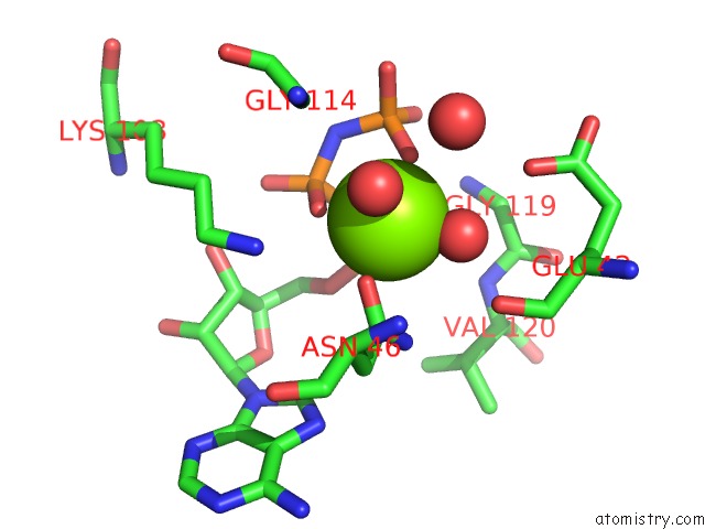
Mono view
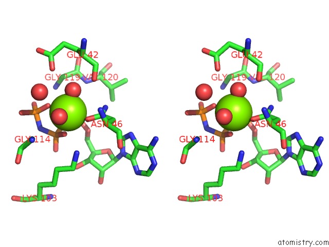
Stereo pair view
Mono view
Stereo pair view
A full contact list of Magnesium with other atoms in the Mg binding
site number 1 of N-Terminal 43 kDa Fragment of the E. Coli Dna Gyrase B Subunit Grown From No Salt Condition within 5.0Å range:
|
Reference:
S.J.Hearnshaw,
T.T.Chung,
C.E.M.Stevenson,
A.Maxwell,
D.M.Lawson.
The Role of Monovalent Cations in the Atpase Reaction of Dna Gyrase Acta Crystallogr.,Sect.D V. 71 996 2015.
ISSN: ESSN 1399-0047
DOI: 10.1107/S1399004715002916
Page generated: Tue Aug 20 13:56:43 2024
ISSN: ESSN 1399-0047
DOI: 10.1107/S1399004715002916
Last articles
Cl in 5WJLCl in 5WJK
Cl in 5WJI
Cl in 5WIV
Cl in 5WJ8
Cl in 5WII
Cl in 5WIU
Cl in 5WGZ
Cl in 5WGY
Cl in 5WHH