Magnesium »
PDB 4wyc-4x7w »
4x59 »
Magnesium in PDB 4x59: Anthranilate Phosphoribosyltransferase Variant P180A From Mycobacterium Tuberculosis in Complex with Prpp and Mg
Enzymatic activity of Anthranilate Phosphoribosyltransferase Variant P180A From Mycobacterium Tuberculosis in Complex with Prpp and Mg
All present enzymatic activity of Anthranilate Phosphoribosyltransferase Variant P180A From Mycobacterium Tuberculosis in Complex with Prpp and Mg:
2.4.2.18;
2.4.2.18;
Protein crystallography data
The structure of Anthranilate Phosphoribosyltransferase Variant P180A From Mycobacterium Tuberculosis in Complex with Prpp and Mg, PDB code: 4x59
was solved by
T.V.M.Cookson,
G.L.Evans,
E.J.Parker,
J.S.Lott,
with X-Ray Crystallography technique. A brief refinement statistics is given in the table below:
| Resolution Low / High (Å) | 45.21 / 1.80 |
| Space group | C 1 2 1 |
| Cell size a, b, c (Å), α, β, γ (°) | 95.234, 78.339, 102.155, 90.00, 111.74, 90.00 |
| R / Rfree (%) | 19 / 22 |
Magnesium Binding Sites:
The binding sites of Magnesium atom in the Anthranilate Phosphoribosyltransferase Variant P180A From Mycobacterium Tuberculosis in Complex with Prpp and Mg
(pdb code 4x59). This binding sites where shown within
5.0 Angstroms radius around Magnesium atom.
In total 2 binding sites of Magnesium where determined in the Anthranilate Phosphoribosyltransferase Variant P180A From Mycobacterium Tuberculosis in Complex with Prpp and Mg, PDB code: 4x59:
Jump to Magnesium binding site number: 1; 2;
In total 2 binding sites of Magnesium where determined in the Anthranilate Phosphoribosyltransferase Variant P180A From Mycobacterium Tuberculosis in Complex with Prpp and Mg, PDB code: 4x59:
Jump to Magnesium binding site number: 1; 2;
Magnesium binding site 1 out of 2 in 4x59
Go back to
Magnesium binding site 1 out
of 2 in the Anthranilate Phosphoribosyltransferase Variant P180A From Mycobacterium Tuberculosis in Complex with Prpp and Mg
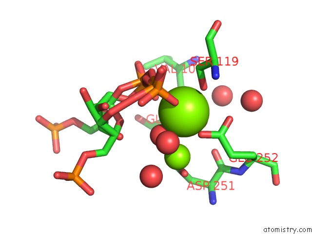
Mono view
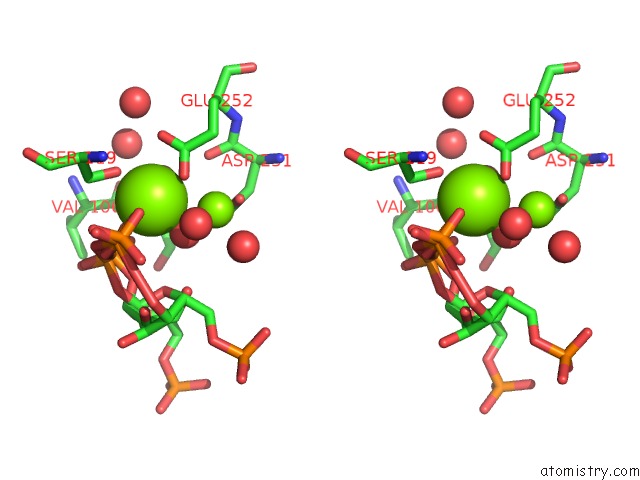
Stereo pair view
Mono view
Stereo pair view
A full contact list of Magnesium with other atoms in the Mg binding
site number 1 of Anthranilate Phosphoribosyltransferase Variant P180A From Mycobacterium Tuberculosis in Complex with Prpp and Mg within 5.0Å range:
|
Magnesium binding site 2 out of 2 in 4x59
Go back to
Magnesium binding site 2 out
of 2 in the Anthranilate Phosphoribosyltransferase Variant P180A From Mycobacterium Tuberculosis in Complex with Prpp and Mg
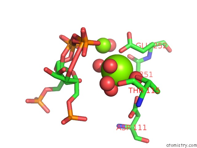
Mono view
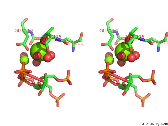
Stereo pair view
Mono view
Stereo pair view
A full contact list of Magnesium with other atoms in the Mg binding
site number 2 of Anthranilate Phosphoribosyltransferase Variant P180A From Mycobacterium Tuberculosis in Complex with Prpp and Mg within 5.0Å range:
|
Reference:
T.V.Cookson,
G.L.Evans,
A.Castell,
E.N.Baker,
J.S.Lott,
E.J.Parker.
Structures of Mycobacterium Tuberculosis Anthranilate Phosphoribosyltransferase Variants Reveal the Conformational Changes That Facilitate Delivery of the Substrate to the Active Site. Biochemistry V. 54 6082 2015.
ISSN: ISSN 0006-2960
PubMed: 26356348
DOI: 10.1021/ACS.BIOCHEM.5B00612
Page generated: Tue Aug 20 14:04:26 2024
ISSN: ISSN 0006-2960
PubMed: 26356348
DOI: 10.1021/ACS.BIOCHEM.5B00612
Last articles
Zn in 9MJ5Zn in 9HNW
Zn in 9G0L
Zn in 9FNE
Zn in 9DZN
Zn in 9E0I
Zn in 9D32
Zn in 9DAK
Zn in 8ZXC
Zn in 8ZUF