Magnesium »
PDB 4xgq-4xsz »
4xj4 »
Magnesium in PDB 4xj4: Crystal Structure of Vibrio Cholerae Dncv 3'-Deoxy Atp Bound Form
Protein crystallography data
The structure of Crystal Structure of Vibrio Cholerae Dncv 3'-Deoxy Atp Bound Form, PDB code: 4xj4
was solved by
K.Kato,
R.Ishii,
R.Ishitani,
O.Nureki,
with X-Ray Crystallography technique. A brief refinement statistics is given in the table below:
| Resolution Low / High (Å) | 47.04 / 1.60 |
| Space group | C 1 2 1 |
| Cell size a, b, c (Å), α, β, γ (°) | 118.090, 51.300, 71.540, 90.00, 93.85, 90.00 |
| R / Rfree (%) | 17.2 / 20 |
Magnesium Binding Sites:
The binding sites of Magnesium atom in the Crystal Structure of Vibrio Cholerae Dncv 3'-Deoxy Atp Bound Form
(pdb code 4xj4). This binding sites where shown within
5.0 Angstroms radius around Magnesium atom.
In total only one binding site of Magnesium was determined in the Crystal Structure of Vibrio Cholerae Dncv 3'-Deoxy Atp Bound Form, PDB code: 4xj4:
In total only one binding site of Magnesium was determined in the Crystal Structure of Vibrio Cholerae Dncv 3'-Deoxy Atp Bound Form, PDB code: 4xj4:
Magnesium binding site 1 out of 1 in 4xj4
Go back to
Magnesium binding site 1 out
of 1 in the Crystal Structure of Vibrio Cholerae Dncv 3'-Deoxy Atp Bound Form
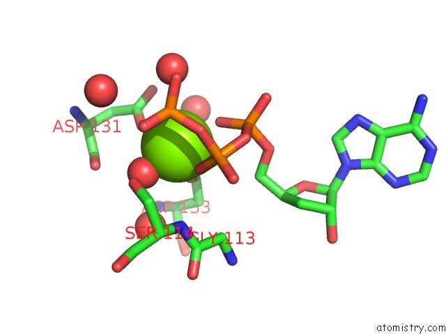
Mono view
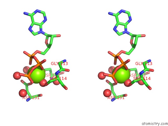
Stereo pair view
Mono view
Stereo pair view
A full contact list of Magnesium with other atoms in the Mg binding
site number 1 of Crystal Structure of Vibrio Cholerae Dncv 3'-Deoxy Atp Bound Form within 5.0Å range:
|
Reference:
K.Kato,
R.Ishii,
S.Hirano,
R.Ishitani,
O.Nureki.
Structural Basis For the Catalytic Mechanism of Dncv, Bacterial Homolog of Cyclic Gmp-Amp Synthase Structure V. 23 843 2015.
ISSN: ISSN 0969-2126
PubMed: 25865248
DOI: 10.1016/J.STR.2015.01.023
Page generated: Tue Aug 20 15:31:58 2024
ISSN: ISSN 0969-2126
PubMed: 25865248
DOI: 10.1016/J.STR.2015.01.023
Last articles
Zn in 9JYWZn in 9IR4
Zn in 9IR3
Zn in 9GMX
Zn in 9GMW
Zn in 9JEJ
Zn in 9ERF
Zn in 9ERE
Zn in 9EGV
Zn in 9EGW