Magnesium »
PDB 4xgr-4xtj »
4xlv »
Magnesium in PDB 4xlv: Crystal Structure of the Activated Insulin Receptor Tyrosine Kinase Dimer
Enzymatic activity of Crystal Structure of the Activated Insulin Receptor Tyrosine Kinase Dimer
All present enzymatic activity of Crystal Structure of the Activated Insulin Receptor Tyrosine Kinase Dimer:
2.7.10.1;
2.7.10.1;
Protein crystallography data
The structure of Crystal Structure of the Activated Insulin Receptor Tyrosine Kinase Dimer, PDB code: 4xlv
was solved by
S.R.Hubbard,
S.Li,
with X-Ray Crystallography technique. A brief refinement statistics is given in the table below:
| Resolution Low / High (Å) | 30.00 / 2.30 |
| Space group | P 32 2 1 |
| Cell size a, b, c (Å), α, β, γ (°) | 66.982, 66.982, 136.580, 90.00, 90.00, 120.00 |
| R / Rfree (%) | 20.4 / 26.6 |
Magnesium Binding Sites:
The binding sites of Magnesium atom in the Crystal Structure of the Activated Insulin Receptor Tyrosine Kinase Dimer
(pdb code 4xlv). This binding sites where shown within
5.0 Angstroms radius around Magnesium atom.
In total 2 binding sites of Magnesium where determined in the Crystal Structure of the Activated Insulin Receptor Tyrosine Kinase Dimer, PDB code: 4xlv:
Jump to Magnesium binding site number: 1; 2;
In total 2 binding sites of Magnesium where determined in the Crystal Structure of the Activated Insulin Receptor Tyrosine Kinase Dimer, PDB code: 4xlv:
Jump to Magnesium binding site number: 1; 2;
Magnesium binding site 1 out of 2 in 4xlv
Go back to
Magnesium binding site 1 out
of 2 in the Crystal Structure of the Activated Insulin Receptor Tyrosine Kinase Dimer
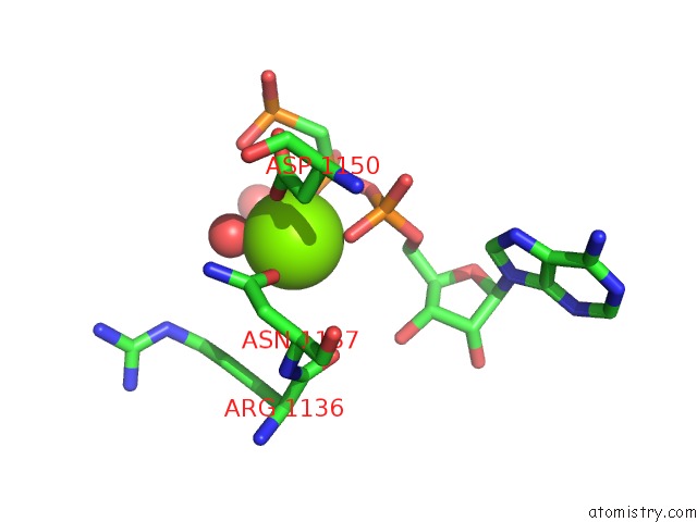
Mono view
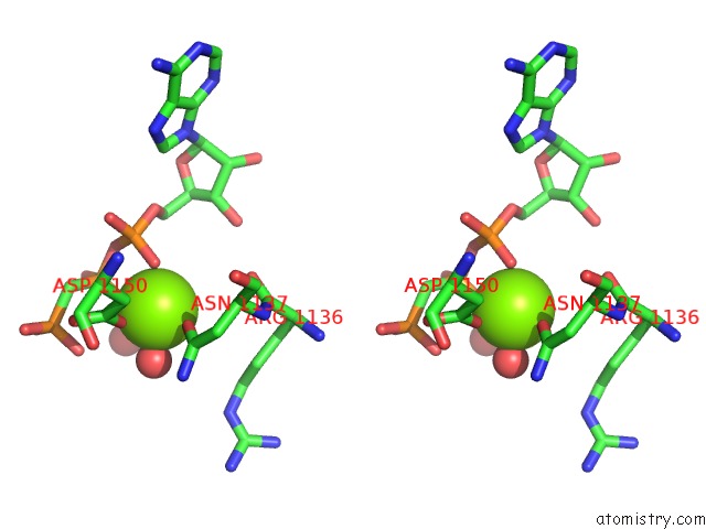
Stereo pair view
Mono view
Stereo pair view
A full contact list of Magnesium with other atoms in the Mg binding
site number 1 of Crystal Structure of the Activated Insulin Receptor Tyrosine Kinase Dimer within 5.0Å range:
|
Magnesium binding site 2 out of 2 in 4xlv
Go back to
Magnesium binding site 2 out
of 2 in the Crystal Structure of the Activated Insulin Receptor Tyrosine Kinase Dimer
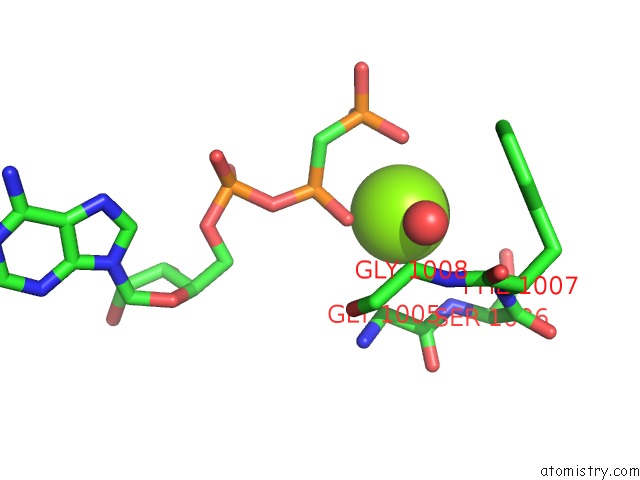
Mono view
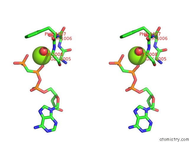
Stereo pair view
Mono view
Stereo pair view
A full contact list of Magnesium with other atoms in the Mg binding
site number 2 of Crystal Structure of the Activated Insulin Receptor Tyrosine Kinase Dimer within 5.0Å range:
|
Reference:
M.Z.Cabail,
S.Li,
E.Lemmon,
M.E.Bowen,
S.R.Hubbard,
W.T.Miller.
The Insulin and IGF1 Receptor Kinase Domains Are Functional Dimers in the Activated State. Nat Commun V. 6 6406 2015.
ISSN: ESSN 2041-1723
PubMed: 25758790
DOI: 10.1038/NCOMMS7406
Page generated: Tue Aug 12 03:09:36 2025
ISSN: ESSN 2041-1723
PubMed: 25758790
DOI: 10.1038/NCOMMS7406
Last articles
Mg in 5G0RMg in 5G5V
Mg in 5G3T
Mg in 5G5T
Mg in 5G5S
Mg in 5G4A
Mg in 5G57
Mg in 5G50
Mg in 5G41
Mg in 5G3Z