Magnesium »
PDB 4xgr-4xtj »
4xsg »
Magnesium in PDB 4xsg: The Complex Structure of C3CER Exoenzyme and Gtp Bound Rhoa (Nadh-Free State)
Protein crystallography data
The structure of The Complex Structure of C3CER Exoenzyme and Gtp Bound Rhoa (Nadh-Free State), PDB code: 4xsg
was solved by
A.Toda,
T.Tsurumura,
T.Yoshida,
H.Tsuge,
with X-Ray Crystallography technique. A brief refinement statistics is given in the table below:
| Resolution Low / High (Å) | 45.29 / 1.80 |
| Space group | P 32 |
| Cell size a, b, c (Å), α, β, γ (°) | 50.745, 50.745, 135.871, 90.00, 90.00, 120.00 |
| R / Rfree (%) | 17.5 / 22.3 |
Magnesium Binding Sites:
The binding sites of Magnesium atom in the The Complex Structure of C3CER Exoenzyme and Gtp Bound Rhoa (Nadh-Free State)
(pdb code 4xsg). This binding sites where shown within
5.0 Angstroms radius around Magnesium atom.
In total only one binding site of Magnesium was determined in the The Complex Structure of C3CER Exoenzyme and Gtp Bound Rhoa (Nadh-Free State), PDB code: 4xsg:
In total only one binding site of Magnesium was determined in the The Complex Structure of C3CER Exoenzyme and Gtp Bound Rhoa (Nadh-Free State), PDB code: 4xsg:
Magnesium binding site 1 out of 1 in 4xsg
Go back to
Magnesium binding site 1 out
of 1 in the The Complex Structure of C3CER Exoenzyme and Gtp Bound Rhoa (Nadh-Free State)

Mono view
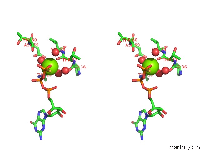
Stereo pair view
Mono view
Stereo pair view
A full contact list of Magnesium with other atoms in the Mg binding
site number 1 of The Complex Structure of C3CER Exoenzyme and Gtp Bound Rhoa (Nadh-Free State) within 5.0Å range:
|
Reference:
A.Toda,
T.Tsurumura,
T.Yoshida,
Y.Tsumori,
H.Tsuge.
Rho Gtpase Recognition By C3 Exoenzyme Based on C3-Rhoa Complex Structure. J.Biol.Chem. V. 290 19423 2015.
ISSN: ESSN 1083-351X
PubMed: 26067270
DOI: 10.1074/JBC.M115.653220
Page generated: Tue Aug 20 15:36:46 2024
ISSN: ESSN 1083-351X
PubMed: 26067270
DOI: 10.1074/JBC.M115.653220
Last articles
Ca in 5UUBCa in 5UUA
Ca in 5UU9
Ca in 5UU8
Ca in 5UN2
Ca in 5UU7
Ca in 5UQZ
Ca in 5ULY
Ca in 5UP7
Ca in 5UN3